Horse Supplements & Horse Products
  | |
| HORSE PRODUCT CATALOGUE | |
| HORSE PRODUCT HARDWARE SPECIAL ORDER CATALOGUE Click here to download a PDF of our Horse Products catalogue in Spanish. |
|
 | |
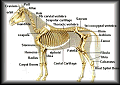
Equine Horse Products Visual | |
| Horse supplement B-VITE crumbles offer 15 mg of Biotin per serving. Keeps in the tradition of good hoof care and a sound stride. Vitamin B6 is added for metabolism of various minerals. Available sizes: 1.13 Kg, 2.5 Kg. | Horse supplement B-VITE PLUS POWDER is Biotin enriched, Offers 20 mg. of Biotin per serving. B-VITE PLUS POWDER will strengthen hooves that are weak and will keep those healthy hooves in peak shape. Also includes: Methionine, Zinc, and Lysine. Available sizes: 1.13 Kg, 2.5 Kg. |
| Specially formulated; high in Sodium and low in Dextrose. Cherry flavoured, ELECTRO-QUENCH supplies electrolytes and minerals that your horse may lose in training. Available sizes:2 Kg, 10 Kg. | A brand of electrolyte in a sodium base, this will help replace body fluids that will be lost during any strenuous activity. The new flavour of Apple will make this is a part of your horses diet. Available sizes: 2.5 Kg, 7 Kg. |
| A formulation of vitamins, minerals, amino acids and electrolytes. Maximizes growth for foals, breeding stallions, promotes bone, hoof, hair and coat development. EQUISUP PELLETS promotes high blood counts and feed efficiency; a complete feed supplement. Available sizes: 2.5 Kg, 7 Kg. | Feed supplement, provides essential vitamins and minerals necessary for growth and development. Available sizes: 2.5 Kg, 7 Kg. |
| Will aid treatment of Lymphangitis, will also help fight the strong scent of horses urine. Available sizes: 500g. | An effective hoof packing for horses. Used for cuts on sheep when shearing, or cuts from wire etc. on any animal. Available sizes: 900 ml. |
| Necessary for normal growth and skeletal muscle levels. VITA B1 is essential for horse that are strenuously exercised. efficiency of B1 may cause appetite loss in horses during times of stress. Available sizes: 1.13 Kg, 7 Kg. | Helps prevent anemia in horses; B12 and Folic Acid are essential for the formation of red blood cells. Available sizes: 1.13 Kg, 7 Kg. |
 Dominion Veterinary Lab Ltd. DVL has a wide spectrum of horse products for the livestock industry. More than one hundred drug formulas continually contribute to the high standards of Canada's, and international agriculture industries. | VITA E AND SELENIUM work hand in hand to promote muscular health and good muscle growth. It is especially important in horses exerting physical effort. Available sizes: 2.5 Kg, 7 Kg.  |
Please Click Here to Contact Us. PHARM-VET HOME PAGE | B-VITE CRUMBLES | B-VITE PLUS POWDER | ELECTRO QUENCH POWDER EQUILECT POWDER | EQUISUP PELLETS | EQUIVIT CRUMBLES |HORSE KIDNEY AND HEMO POWDER PINE TAR | VITA B1 CRUNBLES | VITA B12 WITH FOLIC ACID CRUMBLES VITA E AND SELENIUM CRUMBLES | COMMENTS & QUESTIONS Horse Supplements and Horse Supplies: 3 IN 1® FORMULA Dominion DIN 00391034 Topical Liniment - Tightener - & Brace for Horses Indications: 3 IN 1 FORMULA is a topical liniment used as a tightener and brace with mild antiseptic properties. For use in horses as an aid for temporary relief of leg sprains, swelling, lameness, and muscular soreness. Directions: Shake well before use. ? As a Tightener/Liniment: Relief of an existing condition: Pour a small amount of 3 IN 1 FORMULA into the palm of the hand, apply to affected limbs, and rub briskly until dry. Cover application with cotton and bandage. Apply twice daily. Repeat application until relief of symptoms is noticed, or as directed by a veterinarian. To help prevent leg swelling and muscular soreness: Add 30 mL of 3 IN 1 FORMULA to one litre of warm water. Mix well. Apply once daily to legs after strenuous work. ? As a Body Wash and Brace: Add 30 mL of 3 IN 1 FORMULA to one litre of warm water. Mix well. Apply to entire body or desired area after strenuous work, once daily. This makes a refreshing and stimulating lotion, allowing horses to cool out free of stiffness. ? To Help Relieve Stifle Lameness: Pour a small amount of 3 IN 1 FORMULA into the palm of the hand, apply gently around stifle before a race or workout. Use once daily or as directed by a veterinarian. Caution: For external use only. Apply on a clean leg, from which all other medications have been removed. If condition does not improve after 2 or 3 consecutive applications, or if the condition deteriorates, consult a veterinarian. Warning: Poison - Keep out of reach of children. Active Ingredients: Methyl Salicylate 50.0 mg/ml; Menthol 20.0 mg/ml; Camphor 25.0 mg/ml; Thymol 5.0 mg/ml; Oil of Turpentine (oil of Terebinth) 125.0 mg/ml; Oil of Hemlock 5.0 mg/ml; Oleoresin of Capsicum 0.05 mg/ml. Storage: Store between 15 - 30 degrees Centigrade. Presentation/Packing: 1 Litre bottle; 6 bottles/carton. Disclaimer: Every effort has been made to ensure the accuracy of the information published. However, it remains the responsibility of the readers to familiarize themselves with the horse products information contained on the product label or package insert. NAC No.: 11810012 B.B. JELL-UDDER BALM Indications: B.B. Jell-Udder Balm can be used for all farm animals. It acts as a softening and protective ointment for chapped and damaged udder and teats of cows, mares, and ewes. It can even be used when cowpox are present. B.B. Jell can also be used to lubricate teat dilators and teat tubes. Its soothing and antiseptic ingredients are good for treating all wounds, cuts & abrasions of horses and all livestock. Dosage & Administration: General Use: Apply liberally to the affected area and rub in well. Repeat as necessary Chapped & Damaged Teats: Apply twice daily at milking times. Massage in well. Composition: 2 % Carbolic Acid in an emollient ointment base. Presentation/Packing: 400 gram jar; 12 jars/carton 2 kg jar; 6 jars/carton Keep Out Of Reach Of Children DIN 00250341 DEXAMETHAZONE INJECTION 2 MG Indications: Dexamethazone is an anti-inflammatory agent for use in the treatment of arthritis, snake bite, and ketosis in cattle, as well as supportive therapy for acute diseases and milk fever. For small animals, it is indicated for non-specific dermatitis, supportive therapy following surgery, inflammatory conditions of joints not associated with ruptured ligaments. In horses, it is indicated for carpitis, tendonitis, osselet, myositis, bursitis, sprains, and supportive therapy for fatigue, heat exhaustion & acute infectious diseases. Directions: By intravenous or intramuscular injection. Cattle: 5 To 20 mg Cats: 0.125 To 0.5 mg Dogs: 0.25 To 1.0 mg Horses: 2.5 To 5.0 mg Repeat as necessary. Vary dosage according to the severity of the condition. Contents: Each mL contains: Dexamethazone Sodium Phosphate equivalent to 2 mg Dexamethazone base 1.5 % Benzyl Alcohol Sizes: 100 mL Keep Out Of Reach Of Children DIN 00250325 DEXAMETHASONE POWDER Indications: May be used in those conditions known to respond to cortico-steroid therapy such as certain inflammatory allergic or dermatological conditions. Directions: Cattle & Horses: Use 5 to 10 mg of Dexamethasone base daily, administered orally either by sprinkling on the feed or mixing with a small amount of water and giving as a drench. Contents: Each 15 grams contains: 10 mg Dexamethasone Sizes: 15 g Keep Out Of Reach Of Children DIN 00416010 DUSTING POWDER Indications: Dominion Dusting Powder kills ticks, lice, fleas, and hornflies on cattle, sheep, hogs, and horses; northern fowl mite, chicken mites, lice and fleas on chickens, turkeys, ducks, geese, and domestic game birds. Directions: Beef Cattle, Sheep, Goats, Hogs & Horses: Dust liberally over the entire animal avoiding eyes, and rub thoroughly into the skin. Repeat if necessary, but not more than twice weekly. For control of lice, repeat treatment in 14 to 18 days. Dairy Cattle: Dust carefully along the top line, sides and legs. Do not apply to underline and udder. Repeat if necessary, but not more than twice weekly. Do not apply before milking, only immediately after milking. Wash udders thoroughly before milking. Chickens, Turkeys, Ducks, Geese & Domestic Game Birds: For litter treatment, apply 122 g / square meter. Use a scoop and scatter, or apply by dusting. Avoid contaminating nest, eggs, feeding, and water troughs. For application to birds as a supplement to litter treatment, use 4.5 g per bird. Apply by shaker can or duster. Direct the dust to vent and fluff areas. Repeat in 4 weeks if necessary. Warnings: This control product is to be used only in accordance with the directions on this label. It is an offense under the Pest Control Products Act to use a control product under unsafe conditions. Cautions: Do not use on poultry within 7 days of slaughter. Do not contaminate milking utensils. Harmful if swallowed or inhaled, therefore avoid contact with skin. Avoid breathing dust. Wash after handling. Change contaminated clothing. Contents: 5 % w/w Carbaryl Sizes: 1 kg. Read This Label Before Using Sevin Is A Trademark For The Active Ingredients In This Product. This Product Can Be Used In Any Weather During The Year Keep Out Of Reach Of Children Reg. # 906l MINERAL OIL ELECTROVITE Indications: Electrovite is a multivitamin electrolyte powder for use in livestock under stress conditions. Directions: POULTRY, TURKEYS & PIGS: Dissolve 300 g in 360 litres of water. BEEF CATTLE: (Calves, Bulls, Heifers and Cows) Dissolve 300 grams in the drinking water consumed daily by 60 animals. DAIRY CATTLE: (Cows, Bulls and Calves) Dissolve 300 grams in the drinking water consumed daily by 40 animals. HORSES: Dissolve 1/2 tablespoon in the drinking water consumed daily by each animal. AUTOMATIC MEDIATOR: Dissolve 600 grams in 4.5 litres of water to make a stock solution. Each 30 mL delivery will medicate 4.5 litres. Caution: Prepare fresh solutions daily. Use Electrovite as the only source of drinking water. Contents: Each gram contains: 6610.7·iu Vitamin A 1322.8 iu Vitamin D3 1.98 iu Vitamin E 1.32 mg Vitamin B6 6.6 mcg Vitamin B12 0.165 mg Folic Acid 13.23 mg Ascorbic Acid 3.3 mg Menadione Sodium Bisulfite PLUS AN ELECTROLYTE PREMIX 15 % Magnesium Acetate 10 % Sodium Acetate 10 % Calcium Acetate 15 % Potassium Chloride 10 % Calcium Chloride 20 % Sodium Chloride 20 % Sodium Diacetate Sizes: 300 g & 18 kg Keep Out Of Reach Of Children DIN 00580007 KERAPLEX Indications: As an aid in the treatment of infectious Bovine Keratitis (pink eye) of cattle due to bacteria susceptible to neomycin, and topical as a wound dressing for minor cuts and abrasions of cattle, sheep, and horses. Directions: Hold the container about 15 cm from the site of application, and spray an amount sufficient to cover the eye or cut. Repeat treatment every 6 to 12 hours until improvement occurs. Contents: Each 30 mL contains: 29.6 mg Neomycin Sulfate 71.0 mg Methyl Violet Sizes: 500 mL Keep Out Of Reach Of Children DIN 00486388 KOPPER KARE ‘’NO DANGEROUS GOODS CHARGE’’ FOR SHIPPING Indications: Kopper Kare is indicated for use in the treatment of foot rot on cattle & sheep; thrush, hoof punctures and cracked hooves on horses; ringworm; wounds; after dehorning; as a pad toughener for dogs; and also for spongy hooves. It is a fungicide-antiseptic-astringent liquid. Directions: Remove all dead tissue and cleanse the affected area. Then, use Kopper Kare by squeezing the bottle or applying with a brush or swab. Warning: Avoid contact with eyes and mucus membranes. Keep out of reach of children. Caution: Do not apply to the teats of lactating dairy animals. Not for use on cats or rabbits. Contains: 37.5 % Copper Naphthenate Size: 475 mL DIN 00603481 Indications: Mineral Oil is recommended as an intestinal lubricant to relieve constipation, a dry skin softener, and as a coat dressing. Directions: As A Laxative: Cattle And Horses: 240 to 960 mL depending on body weight and severity of condition. Sheep, Goats & Swine: 60 to 240 mL Dogs & Cats: 5 to 120 mL Repeat dosage daily until condition is corrected. Habitual constipation may be relieved by administering 1 to 2 times weekly. As A Coat Dressing: Mix 1 litre of Alcohol, 1 litre of Water, and 2 litres of Mineral Oil; shake vigorously to make an emulsion. Massage into the coat and skin. Follow with a good brushing. For Use As A Spray: Dissolve 30 grams of soap in 450 mL of hot water. Let cool, then add 450 mL of oil. Finally add 30 mL of this mixture to 180 mL of water for spray. Contents: 100 % Mineral Oil Size: 4 L Keep Out Of Reach Of Children DIN 00097268 NITRO OINTMENT Indications: For use in the treatment of topical bacterial infections of large animals susceptible to nitrofurazone. Directions: Clean area with soap and water. Dry thoroughly, then apply ointment directly to affected area. For a wet dressing, dilute with 2 to 3 parts of sterile water or saline and apply several times daily. Caution: For deep wounds or body cavities, use as directed by a veterinarian. If redness, irritation or swelling persists, consult a veterinarian. Warning: This drug is not to be administered to horses that are to be slaughtered for food. Do not use in food producing animals. Contents: 2 mg Nitrofurazone In a water soluble polyglycol base Sizes: 200 g, 400 g Keep Out Of Reach Of Children DIN 00334308 RACE-VITE PIPERAZINE DIHYDROCHLORIDE 53% PROCAINE PENICILLIN G Indications: A sterile aqueous suspension of Penicillin G Procaine for the treatment of livestock and poultry diseases caused by organisms sensitive to penicillin. Warning: Milk taken from treated animals within 72 hours after the latest treatment must not be used in food. Treated animals must not be slaughtered for use in food for at least 5 days after the latest treatment with this drug. In Cattle: shipping fever syndrome, wound infections, foot rot, bronchitis, bacterial infections associated with pneumonia. In Swine: mastitis, wound infections, bronchitis, tracheitis, bacterial infections associated with pneumonia. In Horses: shipping fever syndrome, strangles, bacterial infection associated with pneumonia, bronchitis, tracheitis. Also in dogs, cats, mink, rabbits, & foxes: otitis externa, bacterial infections associated with pneumonia, bronchitis, tracheitis. Directions: Administer intramuscularly only. In case of anaphylactic or allergic reaction give epinephrine. body weight (kg) minimum dose (mL) 1 - 10 ¼ mL 10 - 20 ¼ - ½ mL 20 - 50 ½ - 1 mL 50 - 100 1 - 2 mL 100 - 200 2 - 4 mL 200 - 400 4 - 8 mL Contents: Each mL contains Penicillin G Procaine 300,000 i.u. with 0.25% w/v Sodium Formaldehyde Sulphoxylate as a preservative. Sizes: 100 mL & 250 mL See Accompanying Insert For Dosage & Directions Keep horse products Refrigerated Store Below 15ºC Shake Well Before Using Keep Out Of Reach Of Children DIN 491020 PHENYLBUTAZONE TABLETS Indications: For oral use as an anti-arthritic, anti-inflammatory. Directions: Horses: 1 to 2 tablets per 225 kg of body weight, not to exceed 4 tablets daily. Reduce dosage as necessary. Contents: 1 g Phenylbutazone Sizes: 100‘s (Prescription Only) Keep Out Of Reach Of Children DIN 00250368 Indications: Piperazine Dihydrochloride is used as a one day wormer, either in water or feed, for the control of Round Worms in poultry, swine and horses. It will also control nodular worms in swine, and small strongyles in horses. Directions: Withholding water is unnecessary. To insure maximum worming of swine withhold water for 12 hours. For best results all medicated feed or water should be consumed the same day. Observe strict sanitation. SWINE: Mix 400 g with 73 kg of feed or 140 L of water. This will treat 125 11 kg pigs, 31 45 kg pigs, 16 91 kg pigs. Treat all swine every 30 45 days starting from 10 weeks. HORSES: Mix 2 teaspoonsful per 45 kg light weight. Add to 45 litres of water per horse. Withhold water 12 hours before treating. CHICKENS: Mix 400 g with 200 kg of feed or 325 litres of water. This will treat 1250 layers or 3100 broilers. Treat all birds every 30 to 60 days starting at 6 weeks. TURKEYS: Mix 400 g with 110 kg of feed or 380 litres of water. This will treat 620 turkeys. Treat all birds every 30 to 60 days starting at 6 weeks. Contents: Each 100 g contains: 53 g Piperazine base (as dihydrochloride) Sizes: 400 g, 2 kg, 10 kg & 50 kg Keep Out Of Reach Of Children DIN 00097128 Indications: Race-Vite is a lyophilized Vitamin B Complex with C. The special two-vial compartment contains a dried portion in one compartment and a liquid diluent in the other compartment. Race-Vite is indicated for the prevention and treatment of Vitamin B Complex deficiencies in large animals. It is designed for addition to solutions of electrolytes and amino acids in the treatment of animals suffering from the effects of stress and dehydration. It may also be given prior to stress producing activity to aid in combating fatigue. Dosage: Administer intramuscularly or by slow intravenous drip according to the following schedule: Horses & Cattle: 10 mL Calves & Colts: 4 to 8 mL Sheep & Swine: 2 to 5 mL Dogs & Cats: 1 mL Composition: Each vial contains: 200 mg Thiamin Hydrochloride (B1) 30 mg Riboflavin (B2) 5-phosphate sodium salt 50 mg Pyridoxine Hydrochloride (B6) 750 mg Niacinamide (B3) 10,000 mcg Cyanocobalamin (B12) 1,000 mg Ascorbic Acid Presentation: 10 mL two-vial compartment Keep Out Of Reach Of Children Din 00419141 SCARLET OIL SPRAY Indications: For use as a dressing for treatment of superficial cuts, wounds, and burns of horses and mules. Directions: Remove pump cap from the top of the container. Direct spray at the site to be treated. Hold the container 10 to 15 cm from the animal and press down on the valve to spray. Apply the spray freely and use it as an open wound treatment or under bandage. The treatment must be repeated once or twice daily as indicated. Caution: In case of deep or puncture wounds or serious burns consult a Veterinarian. If redness, irritation, or swelling, persists or increases, discontinue use and consult a Veterinarian. Contents: Each mL contains: 7.5 mg Menthol 7.5 mg Phenol 7.5 mg Oil of Camphor 7.5 mg Oil of Eucalyptus 7.5 mg Oil of Pine 2.8 mg Oil of Thyme 1.5 mg Peru Balsam 100 ppm Biebrich (Scarlet Red) Sizes: 250 mL spray Keep Out Of Reach Of Children DIN 00526053 SUPER PIP-ZINE 34 SEVIN Indications: Chickens, Ducks, Geese, Turkeys & Domestic Game Birds: Kills northern fowl mite and lice, fleas and fowl ticks. Beef, Dairy Cattle, Sheep, Hogs, Goats & Horses: Kills winter ticks, lice, fleas and hornflies. In & Around Poultry Houses & Farm Buildings: Kills poultry mites, fleas, bedbugs and fowl ticks. Directions: Chickens, Ducks, Geese, Turkeys & Domestic Game Birds: Mix 10 g per litre water and spray at a rate of 45 mL per bird. Repeat in 4 to 8 weeks or as necessary. Beef Cattle, Sheep, Hogs, Goats & Horses: Mix 10 g per litre of water and apply thoroughly to the entire animal at the rate of 4.5 litre per head. Repeat as necessary, but no more than twice weekly. Dairy Cattle: Mix as directed above, but spray carefully along topline, sides and legs. Strictly avoid direct applications to underline and udder. Apply immediately after milking. Do not contaminate milk utensils. Poultry Houses & Farm Buildings: For mites, fleas and bedbugs, mix 15 g per litre water; for fowl tick, mix 40 g per litre of water. Spray 50 to 100 mL per square metre of wall, bedding litter, roost or nest surfaces. Force spray into cracks and crevices. Repeat as needed. Ventilate while spraying. Warnings: This control product is to be used only in accordance with the directions on this label. It is an offense under the Pest Control Products Act to use a control product under unsafe conditions. To prevent residue in milk or meat, do not apply within 1 week of slaughter. Harmful if swallowed or inhaled. Avoid breathing dust or spray mist. Avoid contact with skin. Wash thoroughly after handling. Change contaminated clothing. Sevin is a trademark for the active ingredient in this horse products. Contents: 50 % w/w Carbaryl (1-napthyl-N-methyl carbamate) Sizes: 1 kg, 2 kg & 5 kg Keep Out Of Reach Of Children Reg. No. 9042 Indications: Super Pip-Zine 34 is used for the removal of large round worms in chickens and turkeys; large round worms and nodular worms in hogs; ascarids, some strongyles in horses. Directions: Remove all drinking water the night before treatment. Prepare the amount of medicated water required for one day’s intake. Administer medicated water for one day; then revert to unmedicated water. Treatment every 30 days is recommended. Do not treat brood sows within 30 days of farrowing. CHICKENS: Under six weeks of age use 30 mL in sufficient water to treat approximately 200 birds. Over six weeks of age use 30 mL in sufficient water for every 70 birds. TURKEYS: Under six weeks of age use 30 mL in sufficient water for every 150 poults. Over six weeks of age use 30 mL for every 136 kg. HOGS: Use 30 mL for every 68 kg of body weight. HORSES: Use 30 mL for every 90 kg of body weight. Caution: Consult a Veterinarian before using in severely debilitated animals. Not for the use of dogs and cats. Sizes: 250 & 500 mL, and 1 & 4 L This horse products Is Flavored Keep Out Of Reach Of Children DIN 00097101 DOMINION VETERINARY LABORATORIES LTD. 1199 SANFORD STREET, WINNIPEG, MB, R3E 3A1 Telephone: 204-589-7361 Fax: 204-943-9612 Website: www.domvet.com TOLNOXEQUINE® THRUSH TREATMENT Dominion DIN 02200791 1% w/w Tolnaftate Cream Topical Treatment of Thrush in Horses Tolnoxequine Thrush Treatment is a clean, pleasant smelling cream that applies easily to effectively treat occasional and recurring thrush conditions without sticky messes, stains, or spills. INDICATIONS: For Treatment of Thrush in Horses. DIRECTIONS: Clean the sole and frog of the hoof to remove all dirt and dead tissue. Deposit a layer of cream as it is expressed from the tube, approximately ¼ inch in thickness, along the sulci of the frog. Apply a light wrap or bandage if necessary to keep the cream on the foot. Repeat the cleaning and applications daily for ten days. CAUTION: If the condition worsens or persists beyond 10 days, consult a veterinarian. FOR EXTERNAL USE ONLY. WARNING: Keep out of the reach of children. This drug is not to be administered to horses that are to be slaughtered for food. ACTIVE INGREDIENT: 1% w/w Tolnaftate. STORAGE: Store at Room Temperature. NET CONTENTS: 50 g syringe. Disclaimer: Every effort has been made to ensure the accuracy of the information published. However, it remains the responsibility of the readers to familiarize themselves with the horse products information contained on the horse products label or package insert. NAC No.: 11810832 WART ADE Indications: Wart Ade is a liquid to be applied to warty growths on the body surfaces of horses and cattle. Note: Wart Ade is both non-poisonous and non-caustic when applied externally. Dosage & Administration: Apply to warty growth twice daily, using the dauber supplied. Be sure to cover the entire surface of the wart. Composition: 10 % v/v Oil of Thuja In a fixed oil base Presentation/Packing: 125 mL bottle 12 bottles/carton Keep Out Of Reach Of Children DIN 493597 DOMINION VETERINARY LABORATORIES LTD. 1199 SANFORD STREET, WINNIPEG, MB, R3E 3A1 Telephone: 204-589-7361 Fax: 204-943-9612 Website: www.domvet.com ZEV® Dominion DIN 00549312 An antitussive liquid used to aid in the relief of coughs associated with minor upper respiratory tract problems or mild bronchial irritations in dogs and horses. Indications: For Horses: ? For the temporary relief of coughs associated with minor upper respiratory tract problems or mild bronchial irritations. ? As an appetizer and tonic conditioner. ? For the temporary relief of nasal congestion. For Dogs: ? As an appetizer and tonic conditioner. ? For the temporary relief of coughs associated with minor upper respiratory tract problems or mild bronchial irritations. Directions: Shake well before use. Dosage for Horses: ? For the temporary relief of coughs associated with minor upper respiratory tract problems or mild bronchial irritations: Mix 45 to 60 ml (3 to 4 tablespoons) of liquid ZEV with equal parts of honey, molasses or corn syrup. Place well back on tongue. Administer two to three times daily for 48 hours or as directed by a veterinarian. ? As an appetizer and tonic conditioner: Mix 15 to 30 ml (1 to 2 tablespoons) of liquid ZEV with feed until animal is healthy. To ensure continued health, repeat twice weekly. ? For the temporary relief of nasal congestion: Add 60 ml (4 tablespoons) of liquid ZEV to approximately 10 Litres of boiling water and place where the animal can breathe vapour freely. Dosage for Dogs: ? As an appetizer and tonic conditioner: Mix liquid ZEV with equal parts of honey, molasses or corn syrup. Add this mixture, as indicated below, to feed twice daily until condition improves. ? As a temporary relief of coughs associated with upper respiratory tract problems or mild bronchial irritations: Mix liquid ZEV with equal parts of honey, molasses or corn syrup. Administer this mixture, as indicated below, 3 to 4 times daily for 48 hours or as directed by a veterinarian. Weight of Dog Dosage of Sweetened Mixture(ZEV with equal parts sweetener) 0.5 - 0.9 kg 0.2 - 0.5 ml (5 to 15 drops) 0.9 - 1.4 kg 0.3 - 1.0 ml (10 to 30 drops) 1.4 - 2.3 kg 2.5 - 5.0 ml (½ to 1 teaspoon) 2.3 - 4.5 kg 5.0 - 7.5 ml (1 to 1½ teaspoons) 4.5 - 9.1 kg 7.5 - 10.0 ml (1½ to 2 teaspoons) 9.1 - 13.6 kg 10.0 - 20.0 ml (2 to 4 teaspoons) 13.6 - 22.7 kg 20.0 - 30.0 ml (4 to 6 teaspoons) 22.7 kg & up 30.0 - 40.0 ml (6 to 8 teaspoons) Active Ingredients: Ammonium Carbonate 30.2 mg/ml; Potassium Bicarbonate 52.7 mg/ml; Camphor 0.44 mg/ml; Menthol 4.40 mg/ml; Liquid Extract of Squill 0.50 mg/ml; Solid Extract of Buckthorn 0.70 mg/ml. Warning: Keep out of reach of children. Storage: Store between 15 - 30 degrees Centigrade. Caution: As a cough remedy, if the condition has not improved within 48 hours, consult a veterinarian. Presentation/Packing: 2 Litre jug; 4 jugs/carton 4 Litre jug; 4 jugs/carton Disclaimer: Every effort has been made to ensure the accuracy of the information published. However, it remains the responsibility of the readers to familiarize themselves with the horse products information contained on the horse products label or package insert. NAC No.: 11810904 WHITE LOTION TABLETS Indications: For use as an astringent protectant lotion for abrasions, scratches, and minor raw skin inflammations, primarily of equines. Dosage & Administration: Disperse 2 to 5 tablets in about 1 litre of water and bathe the area for approximately 5 minutes. Composition: Each tablet contains: 1.43 g Zinc Sulfate 1.75 g Calcium Acetate 520 mg Sodium Aluminum Sulfate Presentation/Packing: 100 tablets/jar 12 jars/carton Keep Out Of Reach Of Children DIN 346160 Young horses with both stifles affected may become slightly roach-backed and have difficulties in getting up after laying down. Diagnosis Diagnosis is confirmed by radiographic examination. Treatment Without treatment lameness usually persists. Provided that the lesions are not too severe, surgery is generally successful. SUBCHONDRAL BONE CYST The stifle joint is a common location for subchondral bone cysts, which almost always occur at the point of maximum weight bearing in the lower end of the inside of the femur, the medial femoral condyle. They occur most often in young horses when first starting serious work, but are also found in older horses. Signs The horse shows a sudden onset of a variable degree of lameness. The lameness may be extremely subtle or very severe. The horse usually improves with box rest but the lameness recurs when work is resumed. There is only slight distension of the stifle capsule, if any. Treatment Prolonged rest (6 to 9 months) results in soundness in approximately 60% of horses. In horses which fail to respond to rest surgery can be performed with a fair prognosis. Evidence of partial subchondral bone cyst filling and cartilage repair has been reported in many cases along with increased comfort and accelerated return to performance after using Vet-Stem Cell™Therapy SPRAIN OF THE STIFLE JOINT There is a sudden onset of lameness associated with some swelling of the stifle joint capsule. Diagnosis & Treatment Diagnosis is based on the clinical signs. Rest usually resolves the lameness. The duration of rest depends on the severity of the initial injury. If lameness persists, the stifle should be radiographed. BRUISING OF THE STIFLE REGION Bruising of the stifle region is a common injury either as a result of a kick or hitting a fixed fence when jumping. The latter is a common injury of event horses. There is a variable degree of swelling and lameness. Treatment Anti-inflammatory analgesic drugs are extremely helpful in reducing swelling and removing pain. Plenty of slow exercise prevents the horse from becoming excessively stiff, so if it can be turned out in a small paddock or walked in hand regularly, this is helpful. Provided that there is no underlying bony damage the prognosis is good. If the horse does not improve rapidly the stifle should be examined radiographically. A fracture of the patella or other bony damage might otherwise be missed. Contagious Equine Metritis (CEM, CEMO) is a highly infectious disease of mares caused by Taylorella equigenitalis (previously called Hemophilus equigenitalis). The disease was first diagnosed in 1977 and subsequently spread to many nations. The disease was confirmed in the United States in 1978. Specific regulatory procedures for this disease have been established in the United States and 37 other countries. The organism is carried on the external genitalia of stallions and transmitted at mating to mares, most of which are highly susceptible. The disease is self-limiting and usually clears with sexual rest after about 3 months. Some individuals may take longer to recover and require treatment, and others remain as carriers (harboring the organism but showing no symptoms) for years. Colt foals born to infected mares may be carriers and thus are capable of starting an epidemic by infecting mares during mating. The disease can be the cause of short-term infertility and, very rarely, abortion in mares. SIGNS There is genital inflammation, vaginal discharge and lowered fertility. Affected mares may return to heat unexpectedly, often with shortened interheat periods, but usually breed successfully once the infection has been eliminated. Stallions do not show clinical signs. TREATMENT Most cases resolve without treatment. However, infection appears to persist longer in older mares and recently foaled mares. Treatment is with antibiotics over a 7 to 10 period. Aggressive systemic antibiotic therapy accompanied by routine topical therapy might be required to treat some CEM-positive stallions. PREVENTION Prevention and control of CEM is achievable through a comprehensive programme of breeding farm management that includes early detection and treatment of carrier mares and stallions. Corneal Ulcers follow injuries to the cornea that progress instead of healing; decreased tear production, or entropion. Ulcers which do not heal promptly often become infected. Large ulcers may be visible to the naked eye. They appear as dull spots or depressions on the surface of the corneas. Most ulcers, however, are best seen after the eye has been stained with fluorescein. SYMPTOMS Horses with corneal ulcers often have blepharospasm (tight shutting of the eye in response to the presence of an eye irritant or foreign body on the surface of the cornea), epiphora, and often are extremely sensitive to light (photophobic). These horses may also appear head shy and reluctant to allow physical examination of the head region. TREATMENT Corneal ulcers are dangerous and most receive prompt veterinary attention. Early treatment is vital to avoid serious complications or even loss of the eye. Corneal scrapings examined under the microscope will show if the ulcer is infected. Uncomplicated small surface ulcers respond to topical ophthalmic antibiotic ointment applied 4 times a day. Atropine ointment is used to dilate the pupil. Deep or infected ulcers require intensive antibiotic therapy by subconjunctival injection, or by the intravenous route. Cultures are taken by corneal scraping and antibiotics selected according to the sensitivities. The eylid may have to be sutured together to protect the eye and keep it from drying out. Soft contact lenses also have been used for this purpose. Eye surgery to create a flap of conjunctiva to cover the cornea may be required in difficult cases. White spots of the cornea may persist after healing. If these scars are large enough to interfere with vision, they can be removed by eye surgery. PREVENTION All painful eye disorders should receive immediate veterinary attention. In particular, foreign bodies should be removed as soon as possible to prevent corneal damage. Corticosteroids, which are incorporated into many eye preparations used for treating conjunctivitis and inflamed eyelids, should not be put into eye suspected of having corneal injury. This may lead to rupture of the cornea. Horses wearing fly masks must be checked on a regular basis as they may develop ulcers despite the protection. Unless masks are removed daily, ulcers may go unobserved and progress to a severe stage, before the damage is discovered. As a rule, take masks off at night, to avoid this potential complication. Urinary calculi or stones may form in any part of the equine urinary tract, but the most common site is the bladder (cystic). Calculi are formed by material dissolved in the urine (solutes) being precipitated upon a collection of bladder or other cells, such as red or white blood cells. The factors favoring this precipitation are not well understood but include urine pH—alkalinity increases the formation of carbonate calculi—and the concentration of urine solutes. This can be affected by diet, water intake and loss. The concentration increases when the horse is deprived of water or loses water excessively, as in sweating or diarrhea. If the diet or water fed to a horse has a high mineral content, this also increases solute concentration, while a high-concentrate, low-roughage ratio may allow the deposited solute to cement together more easily. Usually only one calculus occurs at a time, often composed of calcium carbonate. SIGNS All breeds and both sexes are equally likely to develop calculi, athough in mares they become very large before symptoms appear. These are similar to those seen in cases of cystitis, which often is present at the same time. Affected individuals urinate more frequently, with straining and dribbling of urine. Less commonly there may be mild recurrent colic, loss of condition and stilted gait. Occasionally a calculus passes into the male urethra, causing acute obstruction of urine flow. DIAGNOSIS Veterinary help should be always sought in cases in which there is obvious difficulty in passing of urine. Diagnosis of cystic calculi involves urine analysis (the changes are similar to those of cystitis), passage of urinary catheter, and occasionally in the mare, passage of an endoscope into the bladder. TREATMENT Surgical removal of the calculus is the only effective method of treatment. The approach and type of surgery is determined by the size of the stone and the sex of the patient. Some cases may also require treatment for concurrent cystitis. PROGNOSIS Prognosis for cases treated successfully by surgery is guarded because the affected horse may remain predisposed to chronic cystitis and calculus formation. Preventive measure are limited to correct dietary management, particularly in regard to moneral and concentrate proportions, adequate sources of drinking water and prompt veterinary attention to any case of suspected cystitis. In case of osteophyte, there is an abnormal development of bone from surface of joint. Osteophyte is the earliest sign of arthritic change at the margins of the small tarsal joints. The location of the osteophyte formation may vary with the type and/or use of the horse and the stage of the disease. Degenerative joint disease may occur as a result of joint infection (septic arthritis). Most septic arthritis occur fllowing surgery or a puncture wound. Septic arthritis may occur in any joint. Horses with septic arthritis present with severe lameness, joint swelling, fever, loss of appetite, stiffness and pain. Bone changes are more severe and progress rapidly and are usually visible during radiographical examination. Septic arthritis treatment include antibiotics and surgery (in severe cases). Degenerative joint disease may occur as a result of untreated Osteochondritis dissecans (OCD), a disease associated with defective development of bone from cartilage leading to inflammation in affected joints. Often loose fragments of cartilage and/or bone present in joint, usually stifle, hock, fetlock and shoulder. The disease may occur in any joint, but is most commonly seen in the tarsus (hock). Breeds with a high-incidence of tarsal osteochondrosis include Standardbreds, Quarter Horses, Warmbloods and Arabians. Medical reports show that about 76 percent of treated horses raced successfully or performed their intended use following surgery. In many cases, however, additional techniques to improve the healing response in bone and cartilage are needed so as to preserve articular function. The principles of treatment of Degenerative Joint Disease include prevention or treatment of septic arthritis and osteochondritis dissecans; treatment of active soft tissue disease contributing to articular cartilage degeneration, including rest, physical therapy, synovectomy and administration of anti-inflammatory drugs, sodium hyaluronate and polysulfated glycosaminoglycans; treatment of articular cartilage loss or degeneration, including articular cartilage curettage (removal), subchondral bone drilling, and osteophyte removal. The equine practitioner is faced with many choices for controlling inflammation in osteoarthritis (OA.) The proper combination of systemic nonsteroidal anti-inflammatory drugs (NSAIDs), intraarticular steroids, viscosupplementation (injection a preparation of hyaluronic acid into the joint that acts as a lubricant to enable bones to move smoothly over each other and as a shock absorber for joint loads), and chondroprotectants (supplements which work to maintain cartilage health) can be used to treat the disease and stop further progression of degenerative changes to the cartilage surface. Although the whole nutraceutical industry is essentially unregulated, with manufacturers making outrageous claims on horse products that have never been tested at all, are often of poor quality, and occasionally lacking in any active ingredient, ongoing research shows that oral administration of nutraceutical products, such as Glucosamine and chondroitin to the horse is common and easy and is perceived to be a benign treatment for OA in horses. The main goal for use of nutraceuticals is to use them in OA cases to attempt to lower the dose of other drugs that are more problematic while potentially preventing further progress of DJD. Clinical findings have revealed that glucosamine sulfate and chondroitin sulfate are effective and safer alternatives to alleviate symptoms of OA (Glucosamine and chondroitin sulfates in the treatment of osteoarthritis: a survey by de los Reyes GC, Koda RT, Lien EJ). Glucosamine is an amine-sugar that has been marketed as a natural horse products for the treatment of osteoarthritis. It has been popularized in the complementary section of pharmacies as a safe over-the-counter treatment for osteoarthritic pain. Recent research suggests that it may not only provide symptomatic pain relief, but may have a role in chondroprotection. This is due to the fact that articular cartilage is critically dependent upon the regular provision of nutrients (glucose and amino acids), vitamins (particularly vitamin C), and essential trace elements (zinc, magnesium, and copper). Therefore, dietary supplementation programs and nutraceuticals based on antioxidant vitamin C, polyphenols, essential fatty acids, used in conjunction with non-steroidal, anti-inflammatory drugs (NSAIDs) may offer significant benefits to patients with joint disorders, such as OA and OCD. There is also evidence that the combination of oral nutraceutical joint supplements containing high quality glucosamine (GU) and chondroitin sulphate (CS) may be more effective in preventing or treating osteoarthritis in horses than either product alone. Dermatophilosis, also known as mycotic dermatitis, rain scald, mud fever, and greesy heel, is a skin disease caused by fungal organism Dermatophilus congolensis which gains entry to the horse skin when it is saturated by prolonged rain. It is mainly seen in mild wet winters. Symptoms--Lesions are seen on the back, belly and lower limbs. Horses in poor condition and badly cared for at pastures are at risk. Horses with shaggy coats or with feather are particularly at risk. Diagnosis is based on the appearance of the sores and microscopic examination of the organisms. Treatment Dry conditions and improved hygiene should be the first step in therapy. Affected animals must be housed. Long hair shielding the sores must be removed by clipping (sterilizing the blades after use). Astringent lotions are beneficial and antibiotics may be given when the sores are severe. Cracking of the skin may require prolonged careful treatment. Remove long hair and wash with mild soap and tepid water. Areas must be kept dry after initial washing. Dressing with antibiotic ointments is helpful. Rest in a dry area for several weeks will be helpful. Prevention is better than cure. Practise good husbandry, prevent prolonged wetting by providing some shelter, examine regularly for parasites, and never expose a horse or groups of horses to confinement in small muddy paddocks without shelter. Colic refers to abdominal pain, the external signs of which is uneasiness, sweating, biting at the flanks and other signs of pain. The horse gets up and lies down again. Very acute colic may be caused by a twisted gut and generally requires surgery. Studies show that colic may result from congenital (present at birth) malformation of the large colon, enteroliths (pathological formations of mineral concentrations) which cause colonic obstruction, and equine gastric ulcers. There are three main types of colic:tympanitic, spasmodic and obstructive. Tympanitic colic (bloat, gastric tympany, wind colic) Symptoms usually include continuous pain, caused by an overproduction of gas which distends the gut, sweating, a high pulse and a tense abdomen. The horse might take violent attempts to lie down. Frequent attempt to urinate and flatulence is common. This type of colic occurs when a horse is fed large quantitis of grain or coarse mix. Rich green grass grazed in the spring has the same effect. Treatment include analgesics to control pain; anti-spasmodic drugs; vegetable oils to reduce the fermentation process. The treatment of uncomplicated cases is usually successful. Spasmodic colic The most common type of colic, mild in character and short in duration (only a few hours). Seen more often in young horses. Bouts of acute pain, sweating, restlessness, frequent rolling and a tense abdomen are common symptoms. The horse looks repeatedly at its flank. May be caused as a result of blocking the normal passage of gas and intestinal contenets through the gut by migrating strongyle worm larvae; often seen in exhausted, dehydrated horses because of lack of sodium and chlorine ions. Treatment includes anti-spasmodic drugs and sedatives. Obstructive colic This type of colic is further subdivided into obstructive, and less severe impacted types. Obstruction of the stomach and small or large intestine all cause acute pain. The horse rapidly becomes shocked due to the release of toxins into the system. The obstruction can occur on the stomach when it is due to overeating, especially dry material such as nuts, or unsoaked sugar beet. It can also occur in the small intestine as a mechanical obstruction caused by developing tumors, or by a twist, and as a result of hernia. Mechanical obstructions are rare, while food materials are common causes. A sudden change in diet, such as from grass to dry hay or straw, or sand impactions can cause obstruction. Obstructive colic should always be treated as an emergency and is one of the conditions where professional help should be obtained immediately. The decision to operate has to be made quickly, as irreversible changes soon occur in the obstructed tissues. Impacted colic is less obvious in nature. As the impaction builds up, the horse stops eating and passes increasingly dry and scanty feces. The horse spends a lot of time lying down and frequently looks at its flank. Impacted colic responds well to treatment that consists of softening the impacted mass and massage. This may take a few days, but in complicated cases surgery may be necessary. Equine herpesvirus 1 (EHV1) and equine herpesvirus 4 (EHV4) are important equine viruses, causing much damage to the horse industry. EHV1 strains are associated with respiratory disease, abortion, and paralysis, whereas EHV4 strains are primarily associated with respiratory disease. Young horse are at greatest risk to be infected by herpesvirus. The herpesvirus rarely occurs in foals younger than three months of age because they still have their vaccinated mothers' immunity. The great majority of infected foals is seen in sucklings and weanlings between 4 and 12 months of age which develop what veterinarians call "foal snots". The risk of developing the URTD increases with overcrowding, heavy parasite infestation, poor nutritional state, climatic extremes, existing disease, and the intermingling of animals from different social groups. The infection is transmitted by contact with another infected horse and its respiratory discharge. Diagnosis of herpesvirus respiratory disease in horses is performed through laboratory tests. Vaccination of young horses does not prevent the infection, but diminishes the intensity of the disease. The outbreaks of infection are common in large broodmare farm operations, annual yearling sales events and race or show barns in which horses from different places are kept together in enclosed, confined spaces. Two- and three-year-olds usually develop an acute form of herpesvirus URTD with neurological complications and increased risk of abortion. The virus remains in an individual horse over the lifetime and periodically reactivates and transmits from horse to horse and mother to foal. The factors contributing to herpesvirus reactivation include surgery, boarding, prolonged transport, weaning, lactation, extreme weather, and stress. Symptoms of the herpesvirus URTD vary from horse to horse and may range from mild illness to a life-threatening lung inflammation. Most common signs include watery nasal discharge that occurs during the first day of the disease. By the second or third day the nasal discharge often dries to form crust in and around the nostrils and becomes thicker and yellowish. In some horses signs may include loss of appetite (anorexia), fever, lethargy, cough, labored breathing and discharge from the eyes (conjunctivitis). In uncomplicated form of infection, the prognosis for full recovery is usually good and signs usually disappear by the end of second week. With the secondary bacterial infection, primarily by primarily by Streptococcus equi zooepidemicus, the disease is much more severe. Even though the infection by herpesvarus affects mainly the upper respiratory tract, it may lead to other serious complications including abortion, neonatal foal death, brain, lung and eye infections. Treatment includes medications for fever reduction and anti-inflammatory agents for reduction of respiratory tract inflammation and antimicrobials. Horses unwilling to eat or drink may require fluid-electrolyte replacement therapy. Prevention of herpesvirus URTD is the most effective way to control the disease outbreaks. Foals become maximally susceptible to infection by 5 - 6 months of age. Vaccination against EHV-1 and EHV-4 respiratory disease is recommended as part of the preventive, herd-health program for all horses at risk for acquiring infection. Vaccination of young horses does not prevent respiratory infection, but diminishes the intensity of clinical signs and both the magnitude and duration and amount of shedding of infectious virus. Because immunity to EHV-1 and EHV-4 generated by vaccination is of short duration, frequent booster doses are necessary for maximal effectiveness. Equine Infectious Anemia Virus (EIAV) is a lentivirus, of the Retrovirus family with an almost worldwide distribution, infecting equids - horses, mules and donkeys. The EIAV belongs to the family of RNA viruses which generally cause slowly progressive, often fatal diseases. The virus is closely related to the human immunodeficiency virus (HIV), which causes acquired immunodeficiency syndrome (AIDS) in humans. Animals may be acutely or chronically infected. The incubation period is variable, from a matter of days to a few months but generally 1 to 3 weeks. Antibodies usually develop 7 to 14 days after infection and last for life. Infected animals remain carriers of the virus for life. The clinical signs of the acute form of equine infectious anemia are nonspecific; in mild cases, the initial fever may be short lived (often less than 24 hours). As a result, horse owners and veterinarians may not observe this initial sign when a horse is infected with EIAV. These infected horses often recover and continue to move freely in the population. The first indication that a horse was exposed to, and infected with, EIAV may well be a positive result on a routine annual test. The majority of horses are inapparent carriers: they show no visible abnormalities as a result of infection. The inapparent form may become chronic or acute due to severe stress, hard work, or the presence of other diseases. Equine infectious anemia acute form symptoms--These depend upon the stage of the disease. Bleeding, edemas ( excessive accumulation of fluids in body tissues or cavities), rapid breathing, and jaundice occur in the acute disease. During the attack, which usually lasts three to five days, the animal will try to shift its weight from one leg to another because of weakness. It will have shaggy coat and be sluggish. Equine infectious anemia chronic form symptoms--If the horse survives this first acute bout, it may develop enlargement of the liver, spleen and lymph nodes, anemia ( the blood experiences a marked drop in its red corpuscle count and appear thin and watery), fever, different sizes of hemorrhage into the skin (petechia), an irregular heartbeat, and a jugular (located in the region of neck or throat) pulse may become evident. Equine infectious anemia is considered a classic bloodborne infection. People have played an important role in EIAV transmission over the years by using blood–contaminated materials on different horses. The EIAV most frequently is transmitted between horses in close proximity by large biting insects, such as horseflies and deerflies. Control of equine infectious anemia is currently based on detection of anti-EIA virus (EIAV) antibodies through AGID test. However, the current diagnostic methods may fail to diagnose the disease at its early stage. infection. There is no vaccine or treatment for the disease. It is often difficult to differentiate from other fever-producing diseases, including anthrax, influenza, and equine encephalitis. Equine protozoal myeloencephalitis (EPM) is one of the most common neurologic diseases of horses in the United States. It is caused by the protozoa Sarcocystis neurona. The parasite is ingested by the horse through infected food or water, or through direct contact with sporocytes in infected animal feces. Opossums, striped skunks, raccoons, armadillos and cats are intermediate hosts that spread the spores, but horses do not transmit the infection to other animals. The parasite damages the brain, brainstem and spinal cord of the horse. EPM can affect a horse of any age, breed, or sex. Diagnosis--Blood tests detect antibodies to the Sarcocystis neurona but do not indicate if the horse will develop the disease. If antibodies are found in the cerebral spinal fluid along with neurolical signs are positive indicators of the disease. Symptoms--Only a small percentage of horses become sick after ingesting the parasite. Signs include dragging a toe, incoordination, dropped eyelid, malposition of a limb, muscle atrophy, wobbling, head tilt, and occasional lameness. All neurologic disease in horses is not EPM and a complete work-up by your veterinarian is needed in many cases to arrive at a specific diagnosis of the problem. Treatment--Horses are treated for EPM until they have negative test results for antibodies to S. neurona. Many horse develop complications and adverse reactions to medications: fever, loss of appetite, depression, incoordination, mild anemia and abortions. Prognosis--The disease is progressively debilitating to the horse and requires extensive treatment. A vaccine for EPM is available but has unknown efficacy. Response to treatment is an important indicator of survival. Treatment with ponazuril minimizes, but does not eliminate, infection and clinical signs of EPM in horses [1-2]. Prevention--Use good hygiene when it comes to storing a horse's food containers, water buckets and tubs. Equine recurrent uveitis (ERU), also known as periodic ophthalmia or moon blindness, is one of the most common causes of blindness in horses. Classic treatment of ERU includes mydriatics, corticosteroids and nonsteroidal anti-inflammatory drugs. Despite vigorous topical and systemic treatment, however, in many cases, the prognosis for preserving vision remains poor. Symptoms--The signs of this disease are acute pain with the eye closed, discharge, and unwillingness to expose the eye to light. The cornea is cloudy. The eyelids remain tightly shut and tears often run down the cheeks. The conjunctiva and iris are red and inflamed. Recurent attacks diminish the sight until complete blindness results. Causes-- Despite extensive clinical research, the causes of equine recurrent uveitis (ERU) are still unknown. In general, the diseases is thought to be an exaggerated response of the immune system to a wide range of organisms, including leptospires bacteria and and Onchocerca cervicalis parasites. Diagnosis is made based on detailed examination of the eye. Treatment--Long and vigorous treatment is necessary to control the inflammation. Antibiotic treatment is used if a bacterial cause is suspected. Corticosteroids and atropine applied to the eye are also essential. New surgical techniques for ERU have been recently developed giving horses with equine recurrent uveitis a chance of a cure, and the possibility of maintaining their eyesight. The surgery involves making an incision through the sclera and removing the infected base where the organisms are. Then, antibiotics are flushed into that tissue to kill any residual organisms. Any clouded up and inflammatory debris is also removed. Researchers at the ophthalmology department at North Carolina State UniversityWe have developed a drug delivery device (a micro-implant) for treating uveitis. The implant, which delivers a constant amount of cyclosporine (an immunosuppressive agent) within the eye, is made at NCSU and currently is being distributed free of charge to ophthalmologists who request one. PrognosisProper treatment improves the course of the disease, especially if started early. However, the disease is often complicated by secondary catarcts and detachment of retina usually result in irreversible loss of vision. Conventional treatment does not prevent recurrence and horses that have had one attack will almost invariably have another at unpredictable intervals. Prevention—Some eye problems are more preventable than others. Professor Brian Gilger, DVM, MS, Dipl. ACVO, Chief of the Ophthalmology Service, the ophthalmology department at North Carolina State University recommends that horse owners minimize the risk of eye trauma and infection by using a fly mask, and feeding hay on the ground, not from nets, bags, or elevated mangers. "They're horrible about causing eye trauma," he warns. What type of horses are affected most? —Racehorses are the population of horses most often affected by gastric ulcers with an ulcer prevalence between 63 and 90%. In contrast, a much lower prevalence (37%) of stomach ulcers is seen in pleasure horses and the degree of ulceration is less severe. Several endoscopic surveys have indicated that the frequency of gastric ulceration in Thoroughbreds in training is fairly high. Ulcers appear to be chronically progressive during training, but to regress during retirement. Symptoms—Clinical signs that typically are associated with gastric ulceration include recurrent colic for seven or more days, acute colic, poor bodily condition, and/or chronic diarrhea, poor appetite, and weight loss. Often symptoms are less obvious (such as poor performance), or not even noticeable. Foals with ulcers suck only half-hearted, grind their teeth and frequently chew on straw. Causes Large amounts of concentrated high-energy feeds, small rations of forage and a low feeding frequency per day as well as the use of spoiled food can contribute to the development of colics and ulcers. An extended period of diarrhea or treatment with NSAIDs also predisposes to gastrointestinal ulceration. Acids also are the important causes - severe ulceration of the stomach, caused by excess acidity, can develop rapidly in horses deprived of feed or not consuming feed. Compared with being turned out to pasture, stall confinement alone appears to be an important factor in the development of gastric ulcers in horses. Current therapy targets the suppression of gastric hydrochloric acid (HCl) and creation of a suitable environment for ulcer healing. Diagnosis is based on history, clinical signs, gastroscopy, and response to treatment. Treatment—Of the products available, only GastroGard (FDA approved) and ranitidine have been shown to be efficacious in the treatment of EGUS. Ranitidine is often associated with treatment failure as a result of incorrect dosing and lack of owner compliance, because of the three times daily dosing required. Also, EGUS occurs in critically ill neonatal foals, but the underlying cause may be different than in adult horses and acid-suppressive therapy may not be as effective. Other studies demonstrated that omeprazole paste is highly effective in healing gastric ulcers in Thoroughbred racehorses and that the omeprazole paste effectively prevents the recurrence of EGUS. The study also indicates that gastric ulcers in untreated horses did not demonstrate a significant rate of spontaneous healing (contrary to an existing theory of self-healing equine ulcers). Severe ulceration of the gastric squamous epithelial mucosa, caused by excess acidity, can develop rapidly in horses deprived of feed or not consuming feed. Suppression of gastric acidity with the histamine type-2 receptor antagonist ranitidine effectively minimized the area of ulceration caused by feed deprivation. Compared with being turned out to pasture, stall confinement alone appears to be an important factor in the development of gastric ulcers in horses, probably as a result of altered eating behavior. Foal heat, or "ninth day diarrhea" (which actually occurs form days 6 to 14), affects nearly all newborn foals. The stools is soft, pasty-yellow, and not profuse. The foal appears unaffected, remains bright and alert, and nurses at regular intervals. The diarrhea usually lasts fewer than 7 days. Since the diarrhea happens to occur when the mare enters her first heat after foaling, it was believed that hormones in the mare's milk caused the diarrhea. In fact, the same diarrhea also occurs in orphan foals. Newborn foals normally eat manure and feedstuffs such as grain and hay. It appears that the ingestion of these substances may upset the flora of the foal's immature intestinal tract and cause temporary diarrhea. Recent studies show that foal heat diarrhoea is most likely caused by hypersecretion in the small intestinal mucosa (the membrane of the four coats of the intestinal wall), which may overwhelm an immature colon that is unable to compensate by increased fluid and electrolyte (substance that while in solution or its pure state will conduct an electric current by means of the movement of ions; examples of electrolytes: sodium, potassium, chloride, and calcium) absorption. Treatment—Diarrhea of short duration associated with the foal heat requires little treatment. Keep the foal dry and clean around its tail. Zinc oxide ointment is applied to prevent scalding of the buttocks. Luxatives, purgatives and variety of untested equine probiotic products that are commercially available are not recommended as they will make the diarrhea worse. Bacterial Pneumonia--One of the major causes of pneumonia in foals aged between 1 and 6 months with most cases occurring before 4 months is Rhodococcus equi bacteria. Inhalation of the soil-borne organism, Rhodococcus equi, can lead to a chronic and severe pneumonia in young horses and people with weak immune system. In addition, ulcerative colitis is a common result to infection in foals, and dissemination from the lung to other body sites is common in either the horse or man. Rhodococcus equi is largely a soil organism but is widespread in the feces of herbivores (animals that eat only plants). Its growth in soil is considerably improved by simple nutrients it obtains from herbivore manure. About one-third of human patients who have developed R. equi infections had contact in some way with equines or their manure. Others may have acquired infection from contact with soil or wild bird manure. Studies show that foal management practices, environmental management, and preventative health practices are risk factors for development of Rhodococcus equi pneumonia in foals. Housing foals in stalls with dirt floors may increase the risk for development of R equi pneumonia. Also breeding farms with large acreage, a large number of mares and foals, high foal density, and a population of transient mares and foals are at high risk for foals developing pneumonia caused by R equi. Symptoms--The intestinal form of the disease may manifest itself by fever, depression, loss of appetite, weight loss, colic or diarrhea, lameness, nostril flaring and reluctance to move. Heat, pain and severe lameness are characteristics of R. equi septic arthritis. Viral Pneumonia--Equine herpes virus 2 (EHV-2). The EHP-2 is ubiquitous in the general equine population. Foals are born free of EHV-2 infection but virtually all acquire the infection during the first months of life. The common signs of EHV-2 infection include uveitis, nasal discharge, pneumonia and colic. Parasitic Pneumonia--Parasitic pneumonias are not common in horses, especially with today's deworming programs. The typical case involves horses pastured with donkeys infested with the lungworm, Dictyocaulus arnfieldi. In foals, Parascaris equorum is a more common parasite and because its life cycle involves migration through the lung, which can potentially cause signs of respiratory disease. A diagnosis of parasitic pneumonia is often difficult to obtain because it may be difficult to identify larvae in tracheal washes. Parasitic pneumonia could be suspected in foals with a poor deworming history, coughing, nasal discharge and poor response to antimicrobials. Common Symptoms of Foal Pneumonia--The earlier signs of lower airway infection in foals are probably abnormal lung sounds on auscultation, nasal discharge and/or coughing, coughing when they get up or run, respiratory rate above 30 - 40 breaths per minute, or an increased respiratory effort. Crusting may be found at the nostrils or on the bones where the foal may wipe its nose. Nasal discharge, however, may be absent or undetectable if the lower airway discharge is swallowed. Foals with lower respiratory tract infection will often cough while forced to take deep breaths, appear restless or distressed because of the rebreathing bag or show a prolonged recovery after the bag is removed. In severe cases, abdominal breathing, nostril flaring, cyanosis and pronounced exercise intolerance will be present. While most foals with early disease continue to be bright and alert, individuals with severe lesions may be depressed or lethargic and anorexic. Some foals with pneumonia will be reluctant to lie down because of increased breathing difficulties and may become very weak. Treatment--Foals of 1 to 6 months of age will commonly experience respiratory tract infections, which may not necessarily require therapy. However,when labored breathing, fever, depression or appetite, leukocytosis (an abnormal large increase in the number of white blood cells in the blood during an acute infection) occur, antimicrobial therapy should be initiated without delay. Prognosis--A favorable outcome is expected in most cases of foal pneumonia when an appropriate antimicrobial therapy is initiated early in the course of the disease. WHITE LINE DISEASE (Seedy Toe) In this disease the white line disintegrates as the result of infection caused by bacteria, yeast, or fungus. The infection starts at ground level and works its way up the white line to the coronary band. The region commonly affected is the toe back to the quarter. The loss of horn creates a hollow space between the hoof wall and the sole that becomes mealy or "seedy." Eventually, a deep recess, filled with cheesy material and debris, develops between the sole and hoof wall. White line disease seldom occurs in barefoot horses on pasture. Like many other hoof conditions, it is a disease of domestic horse management. The typical horse with white line disease is given limited exercise, bedded in damp wood shavings, kept in a wet stall, and exposed to frequent wet-to-dry episodes such as daily wash-downs or walks in wet grass. TREATMENT. With advanced disease, special shoeing techniques are required. All predisposing conditions should be corrected. CANKER Canker is a chronic infection of the horn tissue of the foot. It begins at the frog and progresses slowly to involve the sole and sometimes the wall. The disease is rare and is found almost exclusively in tropical climates. Canker develops in horses who stand in mud, or in bedding soaked with urine and feces, and who do not receive regular foot care. The cankerous horn tissue of the frog loosens readily, and when removed discloses a foul-smelling, bleeding corium covered with a curdled-white discharge. TREATMENT. consists of moving the horse to a clean, dry stable, or preferable a dry rocky pasture. Remove the shoe and thoroughly clean the frog. Apply a drying agent and bandage the foot to prevent contamination. Consult your veterinarian for best treatment regimen. Because canker often involves the corium, treatment is generally prolonged. THRUSH Thrush is a painful infection involving the frog. It is characterized by a black discharge along with poor growth and degeneration of the horn. The disease can be caused by a number of bacteria, but Fusobacterium necrophorum seems to be the most common. Treat the foot as described for canker. The prognosis is good when the sensitive structures are not involved. Acute gastric dilation is a sudden painful distention of the stomach due to a buildup of fluid or gas. The most commmon cause is grain engorgement. The grain forms a packed mass in the stomach that ferments and draws fluid into itself. Allowing a horse to drink cold water after a heavy workout is another cause of gastric dilation. A secondary type of gastric distention occurs when there is an obstruction in the small intestine or colon. The fecal contents of the bowel back up into the stomach. The stomach progressively enlarges because the horse cannot vomit to relieve the pressure. The pain of acute gastric dilation is severe and violent. It is accompanied by rolling, sweating, kicking at the abdomen, and turning the head as if to bite at the abdomen. Heart and respiratory rates are increased. The horse may exhibit shock with cold extremities. Chronic gastric dilation is a milder condition found in horses who crib and swallow large amounts of air. It also occurs in horses who suffer from gastritis, and those who eat and drink immediately after strenuous exercise. TREATMENT If the horse shows signs of abdominal distress, notify your veterinarian at once. Gastric rupture is a fatal complication of acute gastric dilation and is likely to occur if the distended stomach is not decompressed. Irrigating and flushing the stomach will relieve the problem caused by overeating or over-drinking. Dioctryl sodium sulfosuccinate (DSS) helps to soften a grain impaction. Most horses with gastric dilation are dehydrated and have electrolyte and acid-base imbalances. These are usually corrected by appropriate intavenous therapy. Acute laminitis (founder) may accompany or follow an episode of acute gastric dilation. The equine eye gets its nutrition from fluids that run through special pathways in and out. Obstruction of this outflow of fluid can be the result of an abnormally developed drain (i.e., primary glaucoma) or through damage to the drain from scarring, or accumulation of debris (i.e., secondary glaucoma). The result of this obstruction is retention of fluids and increase in the pressure within the eye. Symptoms include inflammation of the iris, tumors, cataracts, cornea swollen with an excessive accumulation of fluid. At the advanced stages of the disease, the size of the eyeball may increase and eye lenses may dislocate. Equine practitioners usually consider glaucoma as the cause of any unexplained corneal edema or ocular cloudiness and in cases of severe unrelenting ocular inflammation. Accurate measurement with a portable tonometer is essential to make the definitive diagnosis and to monitor the response to therapy. Causes: The most common cause of glaucoma in horses is chronic or recurrent uveitis. Primary glaucoma usually affects both eyes, therefore the unaffected eye usually requires preventive therapy. Secondary glaucoma may not affect both eyes unless the horse has recurrent uveitis in both eyes. Treatment consists in reducing the inflammation using anti-inflammatory medications, decreasing the production of fluids or laser therapy. An outbreak of hives on a shiny coat of a performance horse might seem like an emergency because of the effect it has on plans to show or compete, but in reality it's a potential emergency for another good reason: it signals an ongoing hypersensitivity reaction, one that could escalate to full-blown allergic or anaphylactic shock. Does this mean you you should push the panic button every time your horse gets a case of so-called alfalfa bumps? Yes and no. Most cases of equine hives resolve as quickly as they appear, usually within 24 to 48 hours, and the cause is never figured out. But to shrug them off without a second thought is to invite disaster, because the horse is a practiced and infamous over-reactor to a variety of stimuli, and in most cases of fatal hypersensitivity, the groundwork for the reaction was laid months, maybe even years earlier. Which means that every outbreak of "the bumps" could be a dress rehearsal for the big one. The first bumps usually show up on the side of the neck, followed by the face, the chest, and the upper front legs. They may or may not be itchy. The bumps are initially distinct and steep-walled, and they retain a depression for several seconds when you press on them with your finger (this is called pitting edema). As they grow in size and number, they may coalesce into large plaques of swollen skin. If the outbreak also involves the tissues that line the respiratoty and digestive tracts, there may also be respiratory distress (like a severe asthma attack, with wheezing and an increasingly anxious struggle to get air) and colic pain that leads to diarrhea. The skin condition itself is not dangerous. But it's only a sign of an allergic reaction going on inside the body, and if that allergic reaction is widespread enough to involve the major organ systems such as respiratory and digestive tracts, and it's escalating, the horse could be on a fast track to collapse and death. The biggest mistake is complacency. Because the horse's body tends to be so overreactive, outbreaks of hives are seen pretty often, and as people become acclimated to the condition, they begin to see it as just a nuisance rather than a potential harbinger to a disaster, and it gets "diagnosed" as "alfalfa bumps," spider bites, beestings, creosote allergy, and a number of other unsubstantiated "conditions" that satisfy people's need to pin a reason on things. As a result, the gravity of the situation is completely missed, and the chance to get help early in the process is completely passed by. TREATMENT Medical treatment is aimed at interrupting the allergic response, employing such agents as antihistamines and corticosteroids, but it should be remembered that this is only treating signs. Another thing to bear in mind is that corticosteroids can cause founder. To truly resolve the problem, the underlying allergen must be identified and eliminated. To that effect, some diagnostic tests must be run, including blood tests and skin biopsy. If you notice that your horse is woking harder than usual to get his air, making any wheezing sounds when he breathes, then he is developing a respiratory distress. Loose manure, belly pain or exceptionally gurgly gut sounds are all evidence that your horse might be in serious trouble. Get help. Meanwhile, give him a cool bath and hold ice packs on his swollen eyelids and sooth and help calm his irritated tissues. Do not give him and medications before diagnostic tests. PINWORMS Pinworms (Oxyuris equi) are one cause of the "itchy-tail syndrome." The adult pinworm lives in the large intestine, but doesn't cause much disturbance there. Pinworms deposit their eggs around the anus, causing irritation, tail-rubbing, and hair loss. Eggs can fall to the ground wherever the horse rubs its rear end. Horses are infected with pinworms when they ingest larvae that hatch from eggs several days later. Your veterinarian will do a "scotch-tape prep" to diagnose pinworms, since there are other causes of tail-rubbing. A piece of scotch tape is pressed around the horse's anus, then pressed onto a microscope slide so the tiny pinworms eggs can be examined. Pinworms are easily treated with many dewormers, including the benzimidazole, ivermectin, and the organophosphates. TAPEWORMS Anaplocephala perfoliata and Anaplocephala magna, the equine tapeworms, are less commonly diagnosed than the other parasites. Tapeworms have an interesting life cycle, which involves a stage inside a mite. The mite eats the worm egg in the manure, the horse eats the mite, and the immature tapeworm then emerges and develops into an adult. Adult tapeworms live in and near the horse's cecum, a large dead-end extension between the small and large intestine. Large numbers of tapeworm may cause blockage of the opening into the cecum, ulcers, or even rupture of the intestine. Tapeworms can be difficult to diagnose. A standard fecal egg count may or may not reveal the eggs, and the worms are seldom seen in the manure. Many of the dewormers in use today, including ivermectin, are not effective against tapeworms. Your veterinarian will help you choose the medication to eradicate this parasite. There are many other worms that infest horse, but they probably won't be a problem for you. HABRONEMA larvae live in the stomach wall, where they form tumor like growths. These worms are also the cause of summer sores on the horse's outer body. While the stomach parasite is difficult to diagnose, it is killed by ivermectin; so you are probably eliminating any problem by using this drug in your program. DICTYOCAULUS is a lung worm of horses and donkeys. Infection with lung worms will cause a chronic cough. Immature lung worm larvae are coughed up and swallowed, so they may be seen by microscopic examination of your horse's manure. Ivermectin kills these parasites. FASCIOLA HEPATICA, the liver fluke, is a parasite of cattle and sheep that occasionally infests the horse. Signs include weight loss, colic, and diarrhea. Fluke eggs may be found only with a specific type of fecal examination. Your vet will prescribe specific treatment for flukes since they are not killed with traditional deworming medications. THELAZIA or eye worms are a problem in a few areas. They cause conjunctivitis and irritation of the cornea, which can lead to blindness if untreated. Your veterinarian removes the worms from the conjunctival sac with forceps. Infections of the middle and inner ear can be recognized by signs of labyrinthitis. The labyrinth is a complex organ composed of three semicircular canals: the utricle, saccule, and cochlea. The labyrinth is like a gyroscope. Its purpose is to synchronize eye movements and maintain posture, balance, and coordination. A horse with labyrinthitis will often assume an abnormal posture with a head tilt toward the affected side. Dizziness, incoordination, and loss of balance are evident in the staggering gait, turning and circling toward the affected side, and tendency to lean against walls and fences for support. The horse may exhibit rapid jerking movements of the eyballs, a condition called nystagmus. The usual cause of inflammation of the labyrinth is a bacterial infection of the middle and inner ear. Encephalitis, meningitis, and ryegrass staggers can produce signs of labyrinthitis. These signs can also occur with brain tumors, antibiotic-induced damage to the auditory nerves, antifreeze poisoning, and a condition called idiopathic vestibular syndrome. This syndrome is thought to be caused by a virus. TREATMENT The treatment is directed at the primary disease. Bacterial infection require high-dose antibiotic therapy. The horse should be confined to a quiet, well-bedded stall. Horses that recover from labyrinthitis may exhibit head-bobbing or a coarse tremor of the head, evident during eating or drinking. They are prone to episodes of imbalance and may pose a hazard when used for sport or pleasure. Widespread swelling of a limb below the knee or hock occurs for many reasons, including lymphangitis. A careful clinical examination of both the ffot and the swollen region of the leg is necessary to establish the cause of the swelling. Signs One or more legs are swollen. Hind legs are more commonly affected tha forelegs, and usually the entire leg is swollen up to the level of the stifle, resulting in moderate to severe lameness. Surface lymph vessels are prominent and local lymph nodes are enlarged. Serum may exude through the skin. The rectal temperature may be raised. Heavy-hunter types seem especially prone. Diagnosis The clinical signs are fairly typical. The inflammation is often secondary to a chronic low-grade infection of the leg and careful inspection usually reveals an old wound or wounds. Treatment Treatment aims to eliminate the primary infection and reduce the soft-tissue swelling. Relattively long-term treatment with appropriate antibiotics is combined with cold hosing of the leg, bandaging and exercise. Prognosis Vigorous treatment can produce rewarding results, although the leg may remain prominently thickened. The problem may be recurrent, especially in hind limbs, and careful vigilance is necessary to detect and treat small wounds as early as possible. If your horse has lymphangitis, or is prone to it, never use sports massage. SPORADIC LYMPHANGITIS This non-contagious form of Prognosis normally occurs in horses that have been overfed and kept in with restricted exercise for a few days. Typically the disease would arise in working horses stabled over the weekend. The lymphatic vessels on the inside of the leg are very visible and the leg will be hot and swollen. The horse will stop feeding and exhibit thirst along with patchy sweatting. Constipation is also a coomon feature. Consider using Aconite in the very early stages and Apis or Bovista where the signs fit in later stages when the acute signs have subsided. According to Tim Couzens, author of "Homeopathy for Horses (Threshold Picture Guide)", the most valuable interim remedy to use is Bryonia. ULCERATIVE LYMPHANGITIS This is a mildly contagious form of lymphangitis caused by a variety of bacteria including Corynebacterium pseudotuberculosis, streptococci, and staphylococci. It occurs most frequently in horses kept in poor, unhygienic conditions or where overcrowding occurs and follows wounding or cracked heels. One of the first signs is swelling and pain around the pastern or fetlock joints. Nodules then develop in the tissues, following the course of the lymphatic vessels, especially around the fetlock region. These can grow quite large and burst releasing green pus. Affected areas then ulcerate. Local lymphatic vessels enlarge and become thickened. Affected areas will slowly heal, but more nodules and ulcers can occur over a period of several months. Tim Couzens, author of "Homeopathy for Horses (Threshold Picture Guide)", recommends Merc Sol as the main remedy to use where the nodules have bust and are discharging pus where there are ulcers that bleed easily when touched. Dose: 30c 3 times daily.. Anthracinum is useful where there is no response to Merc Sol, or where crops of discharging nodules keep appearing. Other useful remedies include Muriaticum acid 30c to help heal limb ulcers, Kali iod 30c to prevent more nodules appearing and Silica 30c to help remove scar tissue if the condition has become chronic. Navicular disease is an inflammation of bursa, deep flexor tendon and navicular bone. Bone and tendon develop adhesions which cause pain and lameness. The genetic predisposition to navicular disease is proven nowadays, but otherwise, what causes it is still unclear. Horses with navicular disease have an increased load on the navicular bone in early stance. This has been suggested to be a response to pain in the heel region. The horse responds to heel pain (including pain in the navicular region) by contracting the deep digital flexor muscle to unload the heels. This increases the compressive load on the navicular bone, which may cause damage to the overlying flexor cartilage, which is then painful and identified as navicular disease. Symptoms - Occasional lameness which decreases on rest and after work (horse warms up); standing with affected toe pointed; shuffling gait, especially if both forefeet affected. Diagnosis On X-ray navicular bone has ragged edge and areas of rarefied bone and nerve block. Treatment - Navicular syndrome can be treated in a variety of ways. This is related to the fact that it has a variety of causes. Treatment may include stallrest, non-steroidal anti-inflammatory drugs, and orthopaedic shoeing plus intrabursal injections of short-acting corticosteroids and hyaluronic acid. Many veterinarians recommend shoeing as the primary therapy. Shoeing is performed to correct preexisting problems, enhance physiologic function of the foot, and ease breakover of the foot. Shoeing is reported to be most effective when performed within 8 months of the first signs of lameness. Nonsteroidal anti-inflammatory drugs are not used unless radical changes have been made in the shoeing. Approximately 75% of the horses improve in their performance. If therapy does not improve the horse within 6 to 12 weeks, surgery may give permanent relief. Prognosis - The disease is progressive, and affected horses eventually will need to be retired because of lameness. Often horses with navicular disease remain undiagnosed until the disease has advanced to the stage where problems exist in the foot or to where the conformation-balance of the hoof is irrecoverable. The key to controlling the disease is prevention by maintaining good overall hoof husbandry. Onchocerciasis (Ventral Midline dermatitis) is a skin isease caused by a hair-like worm called Onchocerca cervicalis. The adult worm lives in connective tissue of the horse's neck. The majority of horses in the United States are infected, but a few develop a dermatitis. It is believed that the skin response is due to an allergic reactionn to the dying microfilaria (prelarvae stage of worms released by female worm into the bloodstream). The filaria migrate under the skin and settle primarily on the midline of the abdomen from the chest to the groin, especially around the umbilicus. Other sites are the withers, face, eyelids, and legs. At these sites the parasites produce an itchy skin disorder with redness, moist shallow sores, crusting and scaling, and patchy hair loss. Spots up to 10 inches in diameter can develop. Scarring and loss of skin pigmentation may also develop. These open sores attract Culicoides gnats and other flying insects. Gnats feeding on the open sores pick up filaria and introduce them to a new host. Biting flies and other insects aggravate the skin disorder and create pyoderma. TREATMENT Ivermectin paste is completely effective in ridding the horse of filaria within 2 to 3 weeks. Minor reactions can occur with the use. Veterinary supervision is advised. Adult worms are not affected by deworming agents and therefore serve as a reservoir for recurring infection. To keep the skin free of disease, ivermectin must be repeated at 4-month intervals. A deworming program incorporating ivermectin will effectively control the onchocerciasis. NOTE:The filaria are capable of penetrating the eye and producing uveitis, a leading cause of blindness in horses. Osteochondrosis occurs particularly in young, rapidly growing individuals. There is probably a genetic predisposition in foals. Encouraging a horse to grow quickly using high planes of nutrition may increase the risk of the disease. The stifle and hock are most commonly affected in the hind leg and the shoulder joint in the front leg. The clinical signes are usually evident in the first two years of life. During development of bone, cartilage is converted to bone. Impaired blood supply to the cartilage will delay conversion of the cartilage to bone and result in abnormally thick cartilage on the joint surfaces, the lower layers of which may die. THerefore the cartilage on the joint surface is only loosely attached to the underlying bone and may become detached. This causes inflammation within the joint and production of excess synovial fluid, with the result that the joint capsule becomes distended. Although some joints may have mild osteochondrosis without clinical signs, the majority of lesions cause lameness and deteriorate if not treated. Diagnosis is based on clinical signs and is confirmed by a combination of physical examination, scintigraphy, and radiography. Arthroscopy (examination with a camera inserted into the joint through a small incision of about 1/2 inch) can be used to confirm the diagnosis and treat cartilage and subchondral bone lesions. The disease often affects both stifles, so it is important to take radiographs of both joints even if only one is obviously affected. Symptoms include joint swelling and lameness. Treatment is by surgery. Abnormal cartilage is stripped off the underlying bone and the bone is scraped until healthy bleeding bone is exposed. Prognosis--Young and middle-aged horses with mild osteochondral lesions of the shoulder joints have a good prognosis for return to performance following arthroscopic treatment. Intestinal parasites are an important cause of death in horses of all types. The prevalence of many intestinal helminths is high and, within any population of horses, certain individuals can harbor large numbers of parasites, which may lead to disease. Pinworms (Oxyuris Equi)are nematodes that occur in the large intestine of horse throughout the world. The males are small, but the females may reach 4 inches long and are white-gray in color, with a long tail tapering to a point, hence the name pinworm. SIGNS The presence of the female worms laying their eggs and particularly the presence of the eggs themselves stuck in a gelatinous mass around the anus are irritating, so affected horses will rub their tails. TREATMENT There are several types of deworming compounds: Ivermectin, Moxidectin, Fenbendazole, Pyrantels (Pyrantel Pamoate), and Quasiquantels. Each compound works differently in the horse to destroy internal parasites. Ivermectin paste administered to horses orally continues to be highly effective for treatment and control of a broad range of small and large strongyle species as well as other species of gastrointestinal parasites. PREVENTION Measures are not usually applied specifically for the control of pinworms. Any program of treatment to control the strongyles will control the infection with pinworms. In fact, the presence of pinworms may suggest that the srongyle control program is not being applied properly and the complete worming program should be reviewed. All incoming or returning horses should be quarantine drenched with an effective horse products. This horse products should also be able to remove inhibited (and potentially resistant) larvae. Moxidectin appears to have an advantage here. Ddon't breed from wormy horses. In time this should lead to reduced pasture pollution. Use pasture management. Pick up feces. Pyoderma is a bacterial skin infection that drains pus. Many cases are the result of self-mutilation. When a horse rubs or bites at a persistent irritant to its skin, the skin becomes infected. Always look for another skin disease before concluding that pyoderma is the only problem the horse has. CELLULITIS is an infection of the deep layer of the skin. Most cases are caused by puncture wounds, scratches, and cuts. Horses are aprticularly prone to such injuries. Many wound infections can be prevented by proper early treatment of wounds. Signs of cellulitis are pain (tenderness to pressure), warmth (the skin feels hotter than normal), firmness (not as soft as normal), and change in color (it appears redder than normal). As infection spreads out from the wound, you may feel tender cords which are swollen lymphatic channels. Regional lymph nodes may enlarge. This is a stage beyond cellulitis and is characterized by two disease (ulcerative lymphangitis and malignant edema). A skin ABSCESS is a localized pocket of pus. Pimples, furuncles, and boils are examples of small skin abscesses. An abscess is fluctuant and feels like fluid under pressure. The following skin infections are of particular importance: FOLLICULITIS ("Summer Rash"). This is a hair-pore infection nearly always caused by a Staphylococcus bacteria. It tends to occur in hot weather as a consequence of excessive sweating and friction to the skin from ill-fitting tack. Small pimples appear, usually at points of contact in the saddle or harness areas. These pimples enlarge and form pustules. The pustules rupture and exude pus. Crusts form and the hair becomes matted. Folliculitis can be prevented by good hygiene, such as brushing and cleaning the skin and coat after workouts, and using clean dry blankets beneath saddles. FURUNCULOSIS. This is a deep-seated hair-pore infection with draining tracts and patchy hair loss. It is a progressive form of folliculitis and more difficult to treat. Tail Pyoderma. This condition begins as an itchy skin disorder caused by mange mite or pinworms. As the horse scratches, rubs, and abrades the skin of its tail, secondary staph infection occurs and pustules develop. The ailment is complicated by furunculosis, and by abscesses that rupture and drain in an unending cycle. Hair is lost on the top of the tail. Treatment is most difficult. TREATMENT OF PYODERMA Any underlying itschy skind disorder should be treated to eliminate rubbing, biting, and self-mutilation. Localize the skin infection by clipping away the hair and applying warm soaks for 15 minutes 3 times a day. Saline soaks, made by adding a cup of Epsom salts to a gallon of warm water, make a good poultice. Daily Betadine scrubs help to loosen scabs and promote cleanliness. Topical antibiotics such as nitrofurazone or Triple Antibiotic Ointment are of value and should be applied 2 to 3 times a day. Oral or injectable antibiotics are used to treating wound infections, cellulitis, abscesses, furuncles, and tail pyoderma. Most skin bacteria respond well to penicillin, oxytetracycline, or trimethoprim-sulfadiazine. Rhinopneumonitis virus (rhino) can cause respiratory disease, abortion, or neurological signs. Rhinopneumonitis is caused by an equine herpes virus, EHV-1. The equine herpes virus labeled EHV-2 does not cause disease, and EHV-3 causes a venereal disease). One thing that all herpes viruses have in common is their ability to live within their host indefinitely. Once a horse is infected with rhinopneumonitis, it seems that infection can be reactivated later when the horse is stressed. Young horses are most often affected when they are brought together in groups. Rhinopneumonitis is spread between coughing horses or on the hands of unknowing people. An upper respiratory infection is a common result of virus invasion. Uncomplicated cases will resolve on their own in a week or two. Occasionally the illness will leave the horse susceptible to bacterial pneumonia or a guttural-pouch infection. Another common result of rhino infection is abortion in pregnant mares. Some mares will carry their fetus to term, only to deliver a weak sickly foal with slim chances of survival. The neurological form of rhinopneumonitis is less common. A high fever precedes the development of signs ranging from incoordination and weakness to paralysis. These horses may recover with nursing care, but it can take anywhere from a few days to several months. Two different subtypes of the EHV-1 virus exist in nature. Subtype one usually causes abortion or nervous system disease (myeloencephalopathy), while subtype two (sometimes called EHV-4) is most often the cause of respiratory infections. There are two kinds of vaccine available now to help protect your horse from rhinopneumonitis. One is a modified live-virus vaccine, while the other contains a killed product. Whether killed or live, many rhino vaccines contain only subtype one, the major cause of abortions. Recent studies show that a protection induced by the modified-live virus vaccine is superior to that induced by the inactivated combination vaccine (1). Many vaccines that are currently available contain only EHV-1 subtype one. It's hoped that the two strains of virus have enough in common that vaccination offers some protection against subtype two, the cause of respiratory disease. Vaccines have recently been produced that contain subtype 2, of EHV-4. While respiratory infection is not prevented, its severity is reduced and the length of illness shortened when any vaccine is used. For the competitive show or race horse, any advantage is better than none. Rhino vaccinations are not necessary for the adult pleasure horse. Watch for new developments, since research continues in the search for a better vaccine. Once ingested, the virus multiplies enormously in the cells of the intestine, destroying large numbers of cells that are responsible for the absorption of fluids and nutrients. The resulting diarrhea contains millions of viral particles which contaminate the foaling box and provide a potent source of infection for the next foal that uses the box. Symptoms Foals from 2 days to 6 months may be affected. The first signs include diminished sucking and a foul-smelling diarrhea; this can range from watery to semi-solid and from bright yellow to grey-green in color. Affected foals are dull and depresses but have a normal temperature, young foals can easily become dehydrated and even more depressed with sunken eyes and a tight skin. Diagnosis Diagnosis is made by identification of virus in the feces; several test methods are available. Requesting that the laboratory test specifically for rotavirus, collecting feces early in the course of disease, and sampling several foals improve the chances of viral detection. Treatment Antibiotics are not used. Treatment is generally supportive and requires plenty of fluids. Absorbent drugs and lactobacillus-rich foods such as natural yogurt, help to ensure a swift retuen to normal. A vaccine for pregnant mares to induce colostral antibodies directed at reducing the risk of rotavirus infection in their foals is available. Prognosis The damage done to the intestinal lining is quickly repaired and the diarrhea is normally over in 3 to 4 days. Salmonellosis is a common cause of acute enterocolitis and diarrhea in the horse. However, equine diarrhea can be caused by many bacteria and Salmonella infection accounts for relatively few cases. Horses of all ages and under all conditions of management may be affected although the young, old and debilitated are the most susceptible. There are over 2000 Salmonella types of bacteria, not all of which can cause disease in animals. A small number have been isolated from horses, and Salmonella Typhimurium is by far the most common type identified from clinical disease. Salmonella infection in horses is manifest as a variety of clinical signs. These range from acute diarrhea in young horses; a shock-like condition without diarrhea; mild to severe abdominal pain mimicking equine colic episodes where diarrhea may or may not develop; and protracted diarrhea. Equine salmonellosis is commonly associated with stressful conditions such as transport, exhaustion, general anaesthesia, surgery, antibiotic or deworming medications, change in feed or management or weaning. These events may precipitate changes in food intake, bowel movements and in the normal intestinal microbial flora allowing overgrowth of Salmonella which are present in the intestinal tract of many healthy horses. The development of the disease depends on the immune status of the horse, the bacteria type involved and the presence of external factors. SIGNS Acute diarrhea can occur at any age but is most prevalent in young performance horses. Signs include fever, depression, abdominal pain and dark red mucous membranes. Diarrhea, which may not appear for 2 to 4 days, is watery, foul-smelling, and may persist for up to 4 weeks despite therapy. The shock-like syndrome is more likely to occur in adult horses. Death can follow 6 to 12 hours after the first appearance of abdominal signs. Very young foals may develop an acute generalized infection (septicemia). Older foals may have severe enteritis followed by spreading of the bacteria to joints, the growth region of bones, the lungs, kidneys or central nervous system. Chronic diarrhea is rarely attributed to Salmonella infection but can represent the recovery phase after an acute episode. Confirmation of Salmonella infection depends upon isolating the organism from the feces of affected animals. Horses infected with Salmonella Infantum may develop oral ulcers, limb swelling, weight loss, limb lameness, liver disease, and general weakness. The prognosis is poor. TREATMENT The major objective in treating diarrhea in the horse (whatever the age) is to restore and maintain fluid and electrolyte balance. In the acute stage this may require administration of large volumes of fluids intravenously over 12 to 24 hours or longer. Plasma transfusions may even be indicated in very severe cases. If the animal's condition stabilizes, further fluids can be given orally by stomach tube or by allowing access to water containing electrolyte solutions (fresh water must be available). Normal fecal consistency will be restored most often without using other medications including antidiarrheal agents. Foals with septicemia usually receive a course of IV antibiotics. PREVENTION Salmonellosis is a highly infectious disease, and the build-up of contamination in the environment of the infected horse can place other animals at risk. Ideally, suspected or confirmed cases should be isolated, and strict sanitation measures observed (bleach is the most effective surface disinfectant). Many serotypes of Salmonella Typhimurium are pathogenic for humans. Salmonella Typhimurium definitive type 104 (DT104) has emerged as a common cause of salmonellosis in humans and cattle, yet previous reports involving horses are sparse. The most effective prevention involves good management practices to reduce stressful situations and sanitation. Horses with the history of exposure to antimicrobial drugs prior to hospitalization and abdominal surgery during hospitalization are associated with Salmonella shedding in adult horses with gastrointestinal tract disease. Foals with gastrointestinal tract disease are more likely to shed Salmonella organisms than are adult horses with gastrointestinal tract disease. Risk of Salmonella infections is greater for horses with large colon impactions. Many horses develop superficial sandcracks and never become lame. However, sandcracks can reach into the sensitive layers of the hoof wall or extend into the coronary band, and in those more serious cases, infection can develop resulting in acute lameness. Fissures that originate from the coronary band are generally caused by a defect in the band which results from a wound to that point—the quality of hoof produced at this site is poor, adn when this grows down the hoof a verical crack is formed. Large wounds, bruises or infection breaking out at the coronary band cause a horizontal crack which extends partway round the hoof wall. These rarely cause any trouble until they grow down to near the ground surface, when the section of hoof below the crack may break off. This can cause shoeing problems until the defect has grown out. A permanent sandcrack is formed when the coronary band is damaged. Thus when the cells that produce the horn are damaged they are unable to produce horn to heal up the crack that has occurred. Cracks that extend from the ground surface upwards are caused by an unbalanced or overgrown foot. The unequal forces acting on the foot cause separation of the horn tubules, which extends up the foot until the bond between tubules is stronger than the separating force. Treatment An infected crack must be trimmed out, and all evidence of infection removed. Antibiotic treatment, both local and systemic, will probably be needed, together with daily dressing. Once the horse is sound, the defect can be stabilised by filling the trimmed-out crack with a synthetic resin and fixing it with staples or wire lacing. Another method of fixation is to build up a fiberglass pad over the filled crack. Small screws are used to secure the pad to the hoof wall on each side of the crack. Prognosis Sandcracks oroginating from the ground surface generally respond to regular foot trimming and balancing. Summer Seasonal Recurrent Dermatitis (SSRD), commonly referred to as sweet itch, or summer eczema is considered to be caused by an allergic reaction to the saliva of Culicoides flies (also called midges and "no-see-ums"), grass protein, and filariad worm larvae. Sweet itch occurs only in the late spring and summer when insects are present; during the winter months the skin heals and the hair grows back. The majority of horses do not have an allergic reaction to midge bites, but in an unfortunate 5% the immune system over-reacts producing irritable skin conditions. Horses which are not introduced to the antigens of the biting insects early in life, are most predisposed to develop hypersensitivity to midges saliva. Diagnosis is made based on the history, signs, location of the lesions and response to treatment. Diagnosis is difficult since other allergies and other skin parasites cause similar types of wounds. Symptoms--The fly bites form blisters. Areas of hair loss, crusting, and scaling can be found on the head, neck, shoulders, mane and tail. The allergic reaction to the fly bites starts as a very small, itchy nodule, and the horse will rub and bite at itself. Initially the hair is lost and crusts are seen on the ears and the base of the tail. Eventually the whole back of the horse can become reddened, with crusts and scaly materila over the surface for prolonged periods. Untreated cases develop skin sores and ulcers that are impossible to heal. Treatment & Prognosis--The allergy becomes a chronic condition and requires costant treatment with oral or topical steroids to stop the cycle and anti-inflammatory drugs to control swelling and ease discomfort. Other lotions and remedies have been used, but they will only alleviate the problem temporarily. Moving the horse at least 1/2 mile from the breeding areas od the midges is the key to successful treatment. Prevention--The only effective way to prevent the lesions in hypersensitive horses is to protect them from the bites of Culicoides midges each year. Various management changes can significantly reduce the number of flies and the type of flies: The house fly and stable fly breed in piles of horse manure. Their breeding and numbers can be reduced greatly by effective removal and desinfection of horse manure. Spraying the walls and partitions of the stables with insecticides (every few days to weeks) will kill flies. Spraying the horse with an insecticide. Keeping horses away from water where flies breed will prevent attack on hot days. Use screens and insecticide strips. Use neck straps and head-collar strips or tags with insecticide. Keep horses indoors when the insects feed: from two hours before dusk to two hours after dawn. Keep horses covered with sheets and fly masks. A new vaccine designed to alleviate the distress of sweet itch is available for horse owners to try. The vaccine, which works at removing the excessive reaction to the bites of Culicoides midges, has had promising effects in preliminary trials. Sweet Itch vaccine trial A splint is a soft swelling or a bony enlargement that occurs between the splint bone and cannon bone. The size of a splint can vary from a tiny bump to a large, ugly swelling, and its effects range from an unsightly blemish to a severe lameness. A new, acute, or "hot," splint is usually accompanied by heat, swelling, and pain; the horse may or may not be lame. An old "cold" splint is a hard bump or blemish that usually does not cause pain. CAUSES Splints can have several causes. The syndrome is common in young horses in which the ligament attachment of the splint bone to the cannon bone has not yet hardened into bone. Stretching or tearing of the ligament causes new bone to be laid down, and a firm swelling results. Sometimes, too, the splint bone fractures. Splints in young horses usually occur on the inside of the front legs, since this area bears the most weight. Certain conformation faults predispose horse to developing splints. Horses with "bench knees" (a condition in which the knee is offset to the inside relative to the cannon bone) place more stress on the splint bone. The horse with a base-narrow, toed-out stance is prone to hit the inside of one leg with the opposite foot and a cause of a splint. Splints can occur in horses of any age because of trauma or a blow to the leg. Traumatic splints occur in either the outside or inside of the leg, and are just as common on the hind legs as on the front. Always have your veterinarian radiograph (X-ray) the splint to be sure there is no fracture. Young horses can sometimes continue to work if training is gradual and no fracture is found. However, to reduce the size of the lump, allow the horse to rest and wrap the leg until the splint "cools out" (no heat or pain remains). Alternate cold therapy with sweat wraps to minimize tissue reaction and reduce the size of the bump, since the size of the splint can only diminish while the splint is hot. A permanent blemish often remains after the splint has cooled, but the horse is usually not lame. Large splints can cause lameness even after they are cold if there is enough bone growth to interfere with movement of the ligaments and tendons at the back of the leg. This is less likely to happen if you spot the problem early and take extra precautions to prevent it from progressing. Sporotrichosis is a worldwide disease caused by Sporothrix schenckii, a yeast-like fungus present on vegetation, particularly on the thorns of roses and on plants that carry sharp spicules, or needlelike projections. A draining sore develops at the site of a puncture wound. This usually occurs on the leg but sometimes on the upper body. Nodules appear beneath the skin along the course of lymphatics. The nodules ulcerate, discharge pus, crust over, and heal slowly. The diagnosis is made by taking a sample of wound discharge for fungus culture, or a specimen of infected tissue for fluorescent antibody testing. The disease at times resembles ulcerative Lymphangitis. TREATMENT The frequent application of warm packs is highly beneficial, as the fungus is particularly sensitive to heat. Sporotrichosis often responds to oral iodine therapy when given for several weeks. To prevent relapse, continue treatment 4 weeks beyond healing. In horses that relapse or do not respond to the above, various drugs used to treat human fungus diseases can be tried under close veterinary supervision. Sporotrichosis can be transmitted to people. Use disposable rubber gloves and hygienic techniques when handling infected material. Strangles remains one of the most commonly diagnosed and important infectious diseases of horses world-wide. Strangles infection is extremely contagious. Contaminated tack, feeding utensils, water tubs and bedding are all potent sources of infection. The organism gains entry to the body at the back of the throat and rapidly settles in the tonsils and lymph nodes. From there it spreads to the lymph glands of the head and neck, and causes the swelling and abcess formation so typical of the disease. Most horses recover with no complications, and some 70% develop life-long immunity. It is thought that other 30% become carriers of the disease. Symptoms--Affected animals have a fever, a nasal discharge at first watery then mucous, and poor appetite. Head carriage may be stiffer than normal, swallowing may be difficult and a soft cough heard. Lymph nodes beneath the lower jaw enlarge. These swelling can impair breathing, (hence "strangles") by compressing the airways. Usually horses recover rapidly once the swelling ruptures. Occasionally lymph nodes fail to filter the infection, which spreads to other systems (bastard strangles) producing abcesses, aspiration pneumonia and pleuritis, and even brain damage by inducing metastatic brain abscesses. Diagnosis is made based on clinical signs and confirmed on bacteriological culture, since the presence of swollen lymph nodes under the jaw can indicate many other respiratory diseases (EHV-1 infection etc.) Recent findings suggest that S. equi meningoencephalomyelitis should be considered for foals with neurologic signs that have a history of strangles or exposure to affected horses. Treatment--The nostrils should be cleaned, hot pads applied to the swelling, and soft, easily swallowed feed provided. Strict hygienic measures should be observed as the organism can be transmitted on hands and clothes. Treatment of choice is administration of potassium penicillin and fluids. Antibiotics are used in severe cases, particularly when the infection spreads to lungs and abdomen. Prevention is possible. In a closed herd situation, new arrivals should be isolated for 2 weeks. Since the organism can be shed from draining abcesses for up to 4 weeks and can remain viable for another month or longer, affected horses should be isolated. In-contact animals should be quarantined and observed for signs. However, current antibiotic therapy is often ineffective and thus recent attention has focused on vaccine development. Traditional stringhalt is a poorly understood condition in which the horse hyperflexes one or both hocks. It usually occurs in adult horses, is gradual in onset, and be slowly progressive. The majority of horses recover completely, but a few deteriorate progressively. The condition may be caused by ingestion of a plant toxin. Signs The horse exaggeratedly flexes one or both hind limbs when in motion. The abnormality is sometimes evident at all paces, but it is usually most apparent at walk, especially if the horse is turned or backed, and may disappear at trot. The condition may be intermittent and remain static or deteriorate. Treatment Most often, the cause of the condition is unknown. Some horses are successfully treated by the removal of a piece of the lateral digital extensor tendon at the level of the hock. Prognosis Unless the gait abnormality is extremely severe, affected horses are usually able to perform adequately, including jumping, but are unsuitable fro dressage. A guarded to fair prognosis is warranted after surgery. Some horses improve initially but subsequently relapse. Sometimes stringhalt may develop a complication following trauma to the dorsal metatarsal region. Potential causes include tendon injury that result in abnormal flexion of the tarsocrural joint. Horses may develop stringhalt within 3 months after injury. They can be treated with exercise, including daily hand-walking with pasture turnout, followed by lunging; or surgically, using lateral digital extensor myotenectomy. Of the horses treated with exercise, some may have resolution of stringhalt. Horses treated surgically have varying degrees of improvement, or no change. Strongyles are species of most damaging internal parasites. Strongyles, are divided into two groups, called the large strongyles and the small strongyles. The harmful effects of large strongyles are much greater than of the small ones. The larvae of large strongyles damage blood vessels, while the larvae of small strongyles remains in the wall of the gut. A horse gets infected by ingesting larvae present in grass and forage. Large Strongyles (Bloodworms). One of the three species, Strongyle vulgaris, is the most harmful because its larvae enter the arteries, creates blood clots which block small vessels. This destroys part of the blood supply to bowels and other digestive organs. The larvae live in the arteries for about 5 months, then return to the intestine and develop into adult worms, where they attach to the wall of the bowel. A heavy infection can cause severe anemia Small Strongyles. Small strongyles are the most common internal parasites in adult horses and occur frequently in foals. Larvae produce colic, bleeding and anemia, protein loss, and intestinal malabsorption. PREVENTION OF STRONGYLES. Control of strongyles is most important in all deworming programs. Since the arterial worms are not killed by most dewormers(except for ivermectin), it is extremely important to eliminate the larvae at early stages. An occasional treatment does not offer protection and will not prevent the potentially devastating consequences of these parasites. Most veterinary experts recommend the inclusion of ivermectin in all deworming programs. The summer pneumonia (rattles) usually affects foals aged 2 to 6 months. The respiratory problem is the most common disorder although there may be alimentary tract involvement. Abscesses form in the lungs accompanied by progressive signs of pneumonia. The disease is caused by Rhodococcus equi bacteria (previously known as Corynebacterium equi) that gains entry to the body through inhalation or ingestion. SIGNS Foals rarely show definitive signs until abscesses and bronchopneumonia are well established. Signs include rapid and labored breathing, cough, nasal discharge, a persistent, slightly elevated temperature, poor appetite and weight loss as the disease progresses. The prognosis may be grave in cases with multiple lung abscesses and particularly if the abdomen is involved. TREATMENT Rhodococcus equi is an intracellular organism which provokes an inflammatory reaction. Although the organism is sensitive to several antibiotics and a combination of them have proved successful in some cases, treatment is difficult. Affected foals need prolonged treatment because of the persistence of the bacteria within abscesses in the lung and because immunity to lung infection is poor, the disease tends to recur. The combination of erythromycin and rifampicin have improved the survival of foals infected with R. equi; however, erythromycin can cause adverse reactions in foals and mares, which has prompted the search for alternative therapies. PREVENTION Rhodococcus equi is a robust soil organism widespread in the environment. The organism will potentially multiply wherever there is horse manure. Because the organism reaches the lung by inhalation, dusty manure-contaminated environments (such as are commonly found in loafing paddocks on horse breeding farms in the summer) are potentially lethal sources of infection. It is also important to ensure that loafing paddocks are well grassed, and not totally grazed, reducing them to dusty sandpits.© Copyright 1997 PHARM-VET HORSE PRODUCTS All Rights Reserved. P. O. Box 3506, RPO Redwood CTR, Winnipeg, MB. Canada, R2W 3R4 Telephone: (204) 589-7361/586-3484 Fax: (204) 943-9612  Site Design By Web Wizards | |