
Horse Feed Products
New Products
Raw Material
Antibiotics
Udder Care
First Aid
Wound Treat
Insecticides
Disinfectants
Support Therapy
Nutritional Aids
Links
Antibióticos
Cuidado de la ubre
Primeros auxilios
Tratamiento de heridas
Incecticidas
Disinfectants
Terapia de apoyo
Suplementos nutricionales


|

HORSE SUPPLIES & CATTLE SUPPLIES
SAVE TIME & MONEY
BEST SELLERS
SHOP NOW - CLICK ON ITEM BELOW
 Cattle Supplies Cattle Supplies |
Horse Supplies |
| |
|
| Poultry Supplies |
Hog/Pig Supplies |
| |
|
Order toll-free anytime by phone:
1-800-465-7122
| ABOUT US |
CUSTOMER SERVICE |
| |
|
Over 25 Million Orders Shipped
Click here Equine Vitamin-Mineral Deficiency Chart.
| Shane Chemical-Pharmaceutical National Formulary Supplies Canada |
- Glycerine 4L A clear, colorless, syrupy liquid, used as a humectant and as a solvent for drugs
- Copper Sulfate 400G Used as a vermicide against small nematode worms, germicide, fungicide, viricide, astringent, antiseptic agent and ophthalmic solution. It is also indicated for urethral infection, ring worm, alopecia and wound treatment (caustics).
- Sulfur Powder 2KG Used as an orchard spray and topically as an ectoparasiticide and antifungal in the treatment of skin diseases. Can cause skin irritation in some animals. Called also calcium sulfide, calcium polysulfide, sulfurated lime.
- Borax Powder 400G Used as a herbicide, an insecticide, and a soil sterilant. It is toxic to animals if consumed in moderate to large doses (>0.5 g/kg).
- Citric Acid 400G Is a versatile material and can aid in solving solubility problems for a variety of water administered products in poultry, swine, and other livestock.
- FORMALDEHYDE 500ML Used in the manufacture of resins, particle board, plywood, leather goods, paper, pharmaceuticals and other products.
- ISOPROPYL ALCOHOL 99% 4L Uses include the application as a coolant in beer manufacture, a coupling agent, a dehydrating agent, a polymerization modifier in the production of polyvinyl fluoride, a foam inhibitor, a de-icing agent, a preservative, a heat-exchange medium, and in windscreen wiper concentrates. It is also used as a flavouring agent and in household and personal care products, pharmaceuticals.
- ALUMINUM POTASSIUM SULFATE 2KG Alum is used in water treatment. The addition of alum to raw water causes small particles and colloids to stick together and form heavier particles (floc) which will settle to the bottom. This process is called coagulation or flocculation.
- CALCIUM CARBONATE 400G Antacid. Anti-diarrheal. Supplement.
|
HOME
| GANADO | CABALLOS
| COWS | HORSES
FARM ANIMALS | SHOPPING
CART | CONTACT US | ASK
AN EXPERT
If you cannot find the cattle supplies, horse supplies or livestock supplies you are looking for on our Web
site, or if you have questions, comments or suggestions,
please call us at (204) 589-7361 or Contact Us via e-mail using our online form.
Dominion Veterinary Laboratories guarantees you top quality made-in-Canada products.
For instance, horse supplies, horse drugs, horse medications, horse liniment, horse products, horse supply, horse supplements, cattle supplies, livestock supplies, cattle medicine, cattle prod, cattle feed, cow supplies, livestock antibiotics, and horse medicine.
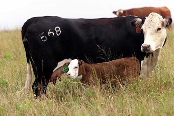
Horse and Cattle Supplies: Calf scours or calf diarrhea causes more financial loss to cow-calf producers than any other disease-related problem they encounter.
But calf scours is not a disease but a symptom of a disease, which can have many causes.
In diarrheas, there is a discharge of more fluid than normal from the bowel, often more frequently than normal.
The discharge can be white, yellow, grey or bloodstained, and is often foul smelling.
Although more common in hand-reared calves, it can also occur in calves which are being suckled by their mothers.
Since a calf is approximately 70 percent water at birth, loss of body fluids through diarrhea can produce rapid dehydration.
Dehydration and the loss of certain electrolytes produce a change in body chemistry in the calf. Although infectious agents may be the cause of primary damage to the intestine, death from scours is usually due to loss of electrolytes, changes in body chemistry, dehydration, and change in acid-base balance rather than by invasion of an infectious agent.
The infectious agent that causes scours can be a virus (BVD, Rotavirus, coronavirus), bacteria (E. coli, salmonella, Enterotoxemia) or protozoa (coccidiosis, cryptosporidium).
Treatment for scours is very similar regardless of the cause.
It should be directed toward correcting the dehydration, acidosis, and electrolyte loss.
Antibiotic treatment can be given simultaneously with the treatment for dehydration. Dehydration can be overcome with simple fluids given by mouth early in the course of the disease.
If dehydration is allowed to continue, intravenous fluid treatment becomes necessary.
The age of the calf when scours begins is an important consideration in its survival.
The younger the calf, the greater the chance of death.
Recent research has indicated that many scour cases can be directly related to lack of colostrum intake by the newborn calf.
A calf that is well mothered and consumes 1 to 2 quarts of colostrum in the first few hours after birth absorbs a higher level of antibodies and is far less susceptible to scours and other calfhood diseases.
Anaplasmosis is a vector-borne, infectious blood disease in cattle caused by the rickesttsial parasites Anaplasma marginale and Anaplasma centrale.
It occurs primarily in warm tropical and subtropical areas.
The disease is not contagious but is transmitted most commonly by ticks.
It can also be transmitted via contaminated needles, dehorning equipment, castrating knives, tattoo instruments, biting flies and mosquitoes.
The intracellular parasite destroys red blood cells.
It causes anemia, fever, weight loss, breathlessness, uncoordinated movements, abortion and death.
Diagnosis is based on clinical signs and the examination of blood under microscope for evidence of the parasite.
Affected cattle either die or begin a recovery within 4 days after the first signs of the disease.
The mortality rate increases with the age of the animal. Unless infected cattle are detected during the early stages of the disease they should not be treated.
If an animal with advanced anaplasmosis is forced to move or becomes excited, it may die from lack of oxygen, also antibiotic treatments do little or nothing to affect the outcome of the disease when given during advanced stages of the disease.
Treatment consists of the administration of tetracycline.
A vaccine is available that helps to reduce the severity of the infection.
If you have any cattle with this disease it is very important to control ticks and follow strict sanitation procedures during vaccinations and other procedures to stop the spread of the disease to healthy animals.
Animals that recover from anaplasmosis are carriers and can spread the disease.
Chlortetracycline also known as CTC can reduce the risk of anaplasmosis.
Chlortetracycline (CTC) consumed at the rate of 0.5 mg / lb. body weight daily during fly and tick season will help to prevent anaplasmosis.
A consistent intake of the correct amount of mineral is crucial to a anaplasmosis prevention program.
CTC is available in medicated feed, free choice salt-mineral mixes or medicated blocks.
Be sure the product is labeled for anaplasmosis control and follow the label instructions exactly.
Anthrax, a highly infectious and fatal disease of mammals and humans, is caused by a relatively large spore-forming rectangular shaped bacterium called Bacillus anthracis.
Most outbreaks occur in areas where animals have previous died of anthrax, as the spores remain viable for decades.
The predominant sign in cattle with anthrax is a progression from a normal appearance to dead in a matter of hours.
Most animals are simply found dead. Once an outbreak begins in the herd animals may be observed with signs of weakness, fever, excitement followed by depression, difficulty breathing, uncoordinated movements and convulsions.
Bloody discharges from the natural body openings as well as edema in different parts of the body are sometimes observed.
After death, the animal's body rapidly decomposes.
Some animals may be saved if treated very early with penicillin or tetracyclines.
Vaccination is very effective in preventing further disease from occurring in animals on a property experiencing an outbreak, however full immunity takes 10 to 14 days to develop.
Antibiotics must not be used at the same time as vaccines are given, since they interfere with the development of immunity.
For animals and humans, anthrax is a reportable disease in the United States.
Local and state health departments, federal animal health officials, and the CDC's National Center for Infectious Diseases should immediately be notified of any suspected cases.
Remember, this is a potentially fatal human pathogen, so appropriate measures must be taken to protect all personnel.
A physician should be contacted for the best preventative measures for all exposed or potentially exposed humans.
Blackleg is a highly fatal disease of young cattle caused by the spore forming, rod shaped, gas producing bacteria Clostridium chauvoei.
The spores of the organism can live in the soil for many years. The bacteria enters the calf by ingestion and then gains entrance to the body through small punctures in the mucous membrane of the digestive tract.
Cattle that are on a high plane of nutrition, rapidly gaining weight and between 6 months and 2 years of age are most susceptible to the disease.
The disease is not transmitted directly from sick animals to healthy animals by mere contact.
The first sign observed is usually lameness, loss of appetite, rapid breathing and the animal is usually depressed and has a high fever. Characteristic swellings develop in the hip, shoulder, chest, back, neck or elsewhere. First the swelling is small, hot and painful.
As the disease progresses, the swelling enlarges and becomes spongy and gaseous. If you press the swelling, gas can be felt under the skin.
The animal usually dies in 12 to 48 hours. In most cases the animal is found dead without being previously observed sick.
The speed with which blackleg kills usually makes individual treatment useless.
Blackleg is almost entirely preventable by vaccination.
The most commonly used clostridial vaccination in cattle is the 7-way type which protects against Clostridium chauveoi (blackleg), Clostridium septicum and Clostridium sordelli (malignant edema), Clostridium novyi (black disease), and three types of Clostridium perfringens (enterotoxemia).
Bloat is a form of severe indigestion marked by a collection of gas in the rumen that the animal is unable to expel.
Normal digestive processes create gases consisting chiefly of carbon dioxide and methane in the rumen. Most of the gases are eliminated by belching.
Gases that are trapped may form a foam or froth in the rumen which further prevents their elimination. Froth formation can be caused by many factors resulting from interactions between the animal, rumen microorganisms, and differences in plant biochemistry.
The main causes of bloat are an inherited tendency for bloat, certain proteins in forage (particularly in legumes), the coarseness of the roughage and the type of rumen microbial population. Pasture bloat usually occurs in animals grazing wheat pasture, lush legumes (alfalfa, Ladino, red clover) or fed green-chopped legumes.
To prevent pasture bloat in cattle you should plant pastures so that no more than 50 percent of the forage mixture is alfalfa or clover, fill cattle on dry roughage or grass pastures before turning to legume pastures, provide grass hay or graze in a rotation using grass pastures.
Visual signs of bloated cattle include distension of the left side of the animal, discomfort as indicated by stomping of feet or kicking of belly, labored breathing, frequent urination and defecation, and sudden collapse.
Brucellosis of cattle, also known as "contagious abortion" and "Bangs disease", is caused by infection with the bacterium Brucella abortus, which can also cause a disease of humans known as "undulant fever". Brucellosis infection of cattle causes abortion or premature calving of recently infected animals, most often between the fifth and eight month of pregancy.
Although federal and state regulations have helped to control this disease, there is still a threat. Infected cows frequently suffer from retained afterbirth, are difficult to get rebred and sometimes become sterile.
Brucellosis is spread from the vaginal discharge of an infected cow or from an aborted fetus. The organism has an affinity for the reproductive tract and abortions, retained placenta, weak calves and infertility frequently occur.
Breeding bulls which are infected, can transmit the disease to cows at the time of service by infected semen. Milk produced front an infected cow may also harbor the organism.
The infected milk creates a public health hazard as this is the organism that causes undulant fever in humans.
There is no treatment for Brucellosis.
Prevention of Brucellosis is accomplished by official calfhood vaccination of heifer calves. Vaccination must be done by an accredited veterinarian at calf ages that vary from two to four months using standard dosage vaccine, or from 4 to 12 months using reduced dosage vaccine.
Each calf must be identified as officially vaccinated in compliance with state and federal regulations. Quarantines are imposed on infected herds by state and federal authorities until the herd has been proven free of the disease.
Bovine Spongioform Encephalopathy (also known as BSE or "mad cow disease") is a progressive degenerative disease that affects to central nervous system of cattle. It belongs to a group of similar but distinct neurological diseases including Creutzfeldt-Jacob disease (CJD) in humans.
The cause of BSE is unknown and there is no known treatment for this fatal disease.
Typically, it takes from two to eight years from the time of infection for the clinical signs of BSE to appear.
BSE-affected animals may display symptoms such as nervousness or aggressive behavior, abnormal posture, lack of coordination and difficulty in rising. When first noticed, clinical signs may resemble those of rabies.
Rabies progresses rapidly over a few days while the symptoms of BSE progress over a period of two to six months. Following the onset of clinical signs, the animal's condition deteriorates until it either dies or is destroyed.
Great Britain's outbreak is believed to have been caused by the inadvertent feeding to cattle of meat and bone meal supplements that were contaminated with an infectious agent. This occurred in the late 1970s and early 1980s.
Once cattle became infected, the BSE agent was recycled in the cattle food chain through the feeding of rendered material from slaughtered animals to other cattle.
This increased the magnitude of the epidemic.
Great Britain banned the feeding of ruminant derived protein (from sheep and cattle) to ruminants in 1988.
the use of potentially contaminated bovine tissue was prohibited in the manufacture of all animal feed. this ban has had an effect, as the number of BSE cases has declined since the winter of 1992-93.
Diagnosis of BSE is based on clinical signs of the live animal followed by the appearance of characteristic lesions in a post-mortem microscopic examination of the brain. There are no tests for the disease in live animals.
BVD (Bovine Virus Diarrhea) infection can cause numerous problems, such as damage to the digestive and immune systems, pneumonia, abortions, calf deformities, and others. Clinical signs in newborn calves infected with BVD include fever, nasal discharge, diarrhea, and inability to move about normally.
Unfavorable reactions frequently follow the use of modified live virus BVD vaccines. The risk of these vaccination reactions should be weighed against the probability of losses resulting from BVD infection before a decision is made about using MLV-BVD vaccines.
Appropriate recommendations should be made by the attending veterinarian after he has assessed the local BVD situation.
Bovine ocular neoplasia includes a variety of benign and malignant skin tumors of the eyeball and eyelids.
Benign tumors are growths that do not spread to other parts of the body and do not tend to grow into surrounding tissues. They can cause local problems with eye function, but do not affect the rest of the body.
Malignant tumors are growths of cells that spread to other parts of the body and tend to invade surrounding tissues. Clearly, it is in the cattlemen's best interest from an economic, humane, and public perception standpoint to treat or market cattle with cancer eye as soon as practical.
Cancer eye appears to affect cattle that have non-pigmented skin, especially around the eye. You can reduce the incidence of cancer eye in your herd by selecting breeding stock with dark pigmentation or color around the eyes and by culling affected animals and their offspring from the breeding herd.
The peak age for cancer eye is between 7 and 8 years of age. It occurs infrequently in cattle less than 3 years of age.
Check eyes whenever cattle are gathered for other routine procedures, especially breeds known to be commonly affected.
Veterinary treatments include surgery, cryosurgery (freezing), hyperthermia (heating), or combinations of these.
The success rate, if treated early, approaches 90 percent.
Given the genetic susceptibility of this condition, you may elect to cull affected cattle rather than treating them.
Cattle with advanced lesions that have spread to other parts of the body or invaded the local tissues around the eye should be humanely destroyed and not transported to market.
If presented, they will be condemned and the presence of cattle with cancer eye at the market could create negative public perceptions.
Coccidiosis continues to be one of the major disease problems for cattle producers.
It is caused by microscopic, one-celled parasites.
Coccidiosis occurs more frequently in calves from one to six months of age, but older cattle, especially those from one to two years, are often affected.
Young calves are usually infected when they are placed in pastures or lots contaminated by older cattle or other infected calves.
Occasionally, mature cattle are infected when they are brought in from large pastures and crowded into small feedlots or barns.
Typical signs of coccidiosis are diarrhea, rough coat, loss of appetite and weight, and general emaciation.
The general weakness may cause the calf to defecate without rising, thus soiling its tail and hindquarters.
In more severe cases the manure may contain blood, mucus, and stringy masses of tissue.
This occurs because the destruction of the epithelial cells results in the sloughing of the epithelium lining the intestine.
Severe straining at defecation may be observed in the more advanced stages.
Death may occur during the acute period, or later from secondary complications, such as pneumonia.
As in many diseases, it is easier to prevent coccidiosis than it is to treat it.
Because several days are required for sporulation, the oocyst stage in manure is the weakest link.
Separating a cow and calf from a contaminated lot interrupts the life cycle and helps control the disease.
Since moisture favors the development of parasites and dryness kills them, practices that reduce the moisture on pasture will decrease parasitic contamination.
Pastures should be well-drained, watering troughs raised above the ground, and grazing should be kept to a minimum on lush grass along the edges of ponds and streams.
In these areas where cattle congregate, overgrazing should be avoided.
Otherwise, animals will be forced to graze to the roots of plants where they may ingest large numbers of parasites.
Segregate severely parasitized animals and treat them with a coccidiostat.
Follow recommended feeding practices.
The effects of parasites are less severe in well-nourished cattle.
Foot-and-Mouth Disease is a severe, highly communicable disease of cattle, pigs, sheep, goats and deer.
It is caused by one of the smallest disease producing viruses known.
There are several different strains of the virus that cause the disease.
The strain now in England and Europe is harder on pigs and cattle but milder in sheep and goats.
Humans do not catch the virus.
The disease is characterized by blister-like lesions on the tongue, nose and lips, in the mouth, on the teats and between the toes which then burst, leaving painful ulcers.
The blisters cause a heavy flow of sticky, foamy saliva that hangs from the mouth.
Infected animals sway from one foot to the other due to the tenderness of the feet.
Although older cattle usually do not die from the infection, they suffer a severe illness which leaves them in a weakened state.
They have high fevers, stop eating, give less milk and become lame.
The virus is extremely contagious and spreads rapidly unless it is contained.
This usually requires quarantining infected farms, followed by slaughtering and burning all susceptible animals.
Anyone having contact with animals in infected countries should not go near susceptible animals for at least five days.
Because the virus is spread so easily, countries with the disease are banned from exporting animals and their products, creating further economic hardship.
Foot-and-Mouth Disease was last seen in the United States in 1929.
The U.
S.
Government places an extremely high priority on keeping the disease out of the country.
Fusobacterium necrophorum and Bacteroides melaninogenicus are the predominant bacteria isolated from footrot. Footrot occurs in cattle of all ages, but it is most common in adults.
The disease is seen year-round, but there is increased incidence in the wet summer and fall months.
Bacteria gain entrance through lesions on the lower part of the foot; the bacteria do not penetrate normal skin.
Anything that can damage the skin between the claws should be considered as predisposing to the disease. Wet manure and mud can soften the skin between the claws and permit infection.
Dried or frozen mud, stones, and stubble can bruise the tissues sufficiently to lower their resistance to disease.
Lameness appears suddenly; usually only one foot is affected.
An animal will put little weight on the affected leg, but will place weight on the limb while walking or running. A moderate fever (103º-104º F.
) may accompany the early signs. The typical early lesion is a break in the skin between the claws.
Pus may be present, but not in large amounts. Edges of the break are covered with necrotic material, and the lesion has a characteristic foul odor.
The foot is swollen and the animal is in acute pain.
Spontaneous recovery may occur, but if the animal is not treated, the lameness may persist for several weeks.
Penicillin, tetracyclines, sodium sulfadimidine, sulfabromomethazine, and other antibacterial agents are used for systemic therapy. Daily treatment begun immediately after onset of lameness usually will give excellent recovery in two to four days.
Treated animals should be maintained on a dry surface until recovered. Recent research has shown that dietary zinc supplementation is effective in treating and preventing footrot in cattle.
Feeding chlortetracycline (CTC) at the rate of 0.5 mg / lb.
body weight daily may also help prevent foot rot. Many mineral mixes and commercial supplements are formulated to contain the correct amount of chlortetracycline (CTC).
Grass tetany is a serious, often fatal metabolic disorder characterized by low levels of magnesium in the blood serum of cattle. It is also called grass staggers and wheat pasture poisoning.
It primarily affects older cows nursing calves less than two months old, but it may also occur in young or dry cows and growing calves. It happens most frequently when cattle are grazing succulent, immature grass and often affects the best cows in the herd.
High nitrogen fertilization reduces magnesium availability, especially on soils high in potassium or aluminum. Grass tetany occurs most frequently in the spring, often following a cool period (temperatures between 45 and 60°F) when grass is growing rapidly, but also is seen in the fall with new growth of cool season grass or wheat pastures.
Typical signs of grass tetany begin with an uncoordinated gait and terminate with convulsions, coma, and death.
Animals on pasture are often found dead without illness having been observed. Evidence of thrashing will usually be apparent around the cow if grass tetany is the cause of death.
The prevention of grass tetany depends largely on avoiding conditions that cause it.
Graze less susceptible animals on high risk pastures. Steers, heifers, dry cows, and cows with calves over 4 months old are less likely to develop tetany. The use of dolomite or high Mg limestone on pastures and including legumes in pasture mixes will decrease the incidence of tetany in grazing cattle.
In areas where tetany frequently occurs, feed cows supplemental magnesium. Supplementation increases blood magnesium levels and alleviates much of the grass tetany problem. Adequate amounts of magnesium must be consumed on a daily basis.
Dominion Veterinary Laboratories guarantees you top quality made-in-Canada products like: horse supplies, horse drugs, horse medications, horse liniment, horse products, horse supply, horse supplements, cattle supplies, livestock supplies, cattle medicine, cattle prod, cattle feed, cow supplies, livestock antibiotics, and horse medicine. Infectious bovine rhinotracheitis (commonly called IBR or red nose) is an acute, contagious virus disease of cattle. Often implicated as an infection which initiates the shipping fever complex.
This infection usually occurs in the air passages of the head and the wind pipe. However, in females this virus also causes inflammation of the vulva and vagina and abortion. Abortion occurs about 20 to 45 days after infection.
Cattle of all ages that have not been vaccinated or have not recovered from the disease are susceptible to IBR. The use of modified live vaccines on non-immune pregnant cows or on animals in contact with pregnant cows could possibly cause abortion. An intranasal vaccine is available that can be used on pregnant cows, if necessary.
It is advisable that heifers be vaccinated or revaccinated 30 to 60 days before breeding.
Many IBR vaccines include the entire IBR/PI3/BRSV/BVD shipping fever complex.
Horn flies, face flies, stable flies, ticks, lice and mites are the major external parasites in beef cattle.
Horn Flies are about half the size of house flies and are dark gray. They are blood-sucking flies that stay on the shoulders and backs of cattle almost continuously. During extremely hot weather or when it rains, they may move to the protected underside of the animal.
When disturbed, horn flies will fly up in a swarm but they will return to the animals almost immediately. A horn fly leaves the back of a cow or calf only to lay eggs in fresh manure. They suck blood from the host 24 hours a day. Individual flies pierce the skin with their short, tube-like mouthparts 20 to 30 times per day to ingest a small amount of blood. Their feeding activity is painful and annoys the animals, as well as causing some blood loss.
There are many effective options to keep horn fly numbers below the 100 fly per animal treatment threshold. Cost, convenience, and herd management practices, such as grazing rotation, can be considered when designing a control program that fits best. Backrubbers allow cattle to treat themselves while loafing and scratching. Dust bags are most effective when used where cattle have to pass under them daily to get to water or mineral.
Feed additives target horn fly maggots breeding in fresh animal manure. High pressure sprays can be used to treat cattle thoroughly and inexpensively on a per head basis. An insecticide bolus is a large pill-like formulation that is given to the animal with a standard balling gun. Insecticide-impregnated cattle ear tags release small amounts of an insecticide which are distributed over the animal during grooming or rubbing.
Pour on insecticides are ready-to use formulations that are applied in measured doses to animals based upon body weight.
Face Flies closely resemble house flies. Face flies cluster on the faces of cattle and feed on secretions from the mucus membranes of the eyes, nose, and lips. Face flies do not suck blood. They do irritate the surface of the eyeball and carry and spread bacteria and viruses that contribute to pinkeye problems.
They spend only a small portion of their life on cattle which makes them more difficult to control than horn flies.
Stable flies are sometimes called biting house flies. The look very much like house flies. They feed primarily on legs and lower abdomen of cattle.
The mouth parts penetrate the skin and allow them to engorge on blood two to three times a day depending on the weather. Once full they move to a resting place, usually in the shade, to digest the blood meal. The blood loss and pain associated with the bite of stable flies results in substantial economic loss.
Ticks cause blood loss, discomfort, and spread diseases like anaplasmosis.
Tick control is extremely difficult in areas with high tick populations. High concentrations of ticks usually occur in brushy pastures and woodlands so habitat management is an important part of tick control. Control on cattle through persistent use of approved pesticides is achieved by spraying, dipping, ear tags, pour-ons, dust, and backrubs. A good residual insecticide is necessary to prevent infestation.
Lice cause skin irritation and itching. Both biting and sucking lice infest cattle. Infested cattle can experience reduced appetite and appear unthrifty.
Lice reside entirely on the host cow.
Lice are present on cattle year around but increase in numbers in winter. In spring most parasites are lost with the winter hair coat. Lice control is most important in the fall and early winter when the lice populations increase. Treat with approved products.
Treatment needs to be repeated in three weeks to kill hatching lice since most insecticides do not successfully kill eggs. Sprays and pour-ons are common methods to treat cattle lice.
Mite infestation is called mange in cattle. A serious form of mange is called scabies.
Scabies is caused by sarcoptic and psoroptic mites and must be reported to the disease control authorities. A less severe mange is caused by chorioptes, demodex, or psorergates mites. Mites are spread through close contact. Cattle infested with mites suffer hair loss and a thickening of the skin.
Severe infestations can weaken cattle and make them vulnerable to diseases. Scabies can result in severely debilitated animal. Control of mites is difficult because mites burrow into hide. Injectable products or pour-on products with systemic activity work to control mites best.
As with lice, a second application is necessary in two to three weeks to kill newly hatched mites.
The economic losses from worm parasite infections of cattle can be significant. Calves under one year of age are more susceptible than older cattle who frequently have been exposed to the parasites and have developed a degree of immunity.
Adult worms in cattle produce eggs that are passed in the manure.
The eggs hatch, producing larvae that develop and move up onto the pasture grasses where cattle consume them. Eggs can survive the winter and hatch out with warm weather. Infection is most likely to occur when temperatures are between 60° and 80°F. and there is adequate rainfall.
Deworming prior to the grazing season will greatly reduce the contamination of pastures during the grazing season. Cows dewormed in the fall usually have a higher conception rate the next breeding season, winter better and wean heavier calves.
There are several several anthelmintics approved for use in beef cattle. It is probably a good idea to rotate the wormer you use.
Consult your veterinarian concerning the type to use and the timing to be the most cost effective for your area. Several are listed below by active ingredient and (brand).
Albendazole (Valbazen) is available in paste or suspension. It is effective against all intestinal worms including tapeworms, and lungworms as well as liver flukes.
It has a 27-day withdrawal for slaughter. It should not be used in animals during the first 45 days of pregnancy.
Fenbendazole (Panacur, Safeguard) is available as a stable suspension or granules. It is effective against roundworms in the gut, larval forms in the tissues, and lungworms.
Withdrawal time to slaughter is 8 days.
Ivermectin (Ivomec) for cattle is an effective medication against the internal worm parasites including lungworms as well as cattle grubs and sucking lice. It is available in injectable or pour-on formulations. Withdrawal time to slaughter is 35 days.
Levamisole (Levisol, Tramisol) is available in boluses, a paste for oral administration, as a pour-on or an injectable form. Levamisole is effective against roundworms and lungworms. Withdrawal time is orally 2 days and injected 7 days.
Morantel tartrate (Rumatel) comes in boluses or crumbles for oral use.
It is effective against roundworms, and has a 14-day withdrawal time to slaughter.
Oxfendazole (Synonthic) is a new wormer that is effective against intestinal parasites including tapeworms. This wormer has a unique delivery system in that the wormer is injected directly into the rumen. Oxfendazole is also available in the drench form.
It has a 7 day withdrawal time to slaughter.
Thiabendazole (Omnizole, TBZ) for oral administration is available in paste, boluses, suspension, or crumbles. It is effective against roundworms. Withdrawal time to slaughter is 3 days.
Johne's Disease (pronounced YO-knees), or paratuberculosis, is a chronic wasting disease that causes considerable production losses in adult cattle, sheep, goats, deer, llamas, elk, and bison, and other ruminants. The disease is caused by Mycobacterium paratuberculosis, a bacterium related to tuberculosis.
Johne's disease typically starts as an infection in calves, though visible signs do not generally appear until cattle are 2 to 5 years of age (and sometimes much older). The infection is difficult to detect in its early stages. This bacterium causes an inflamed intestinal tract that results in severe weight loss and diarrhea and lower milk production. Infected cattle frequently eat well, and look bright, however, they appear to be unthrifty. Body temperature may or may not be elevated. There is no cure for Johne's disease once an animal becomes infected.
Eradication of Johne's disease is extremely difficult because of its insidious nature, long incubation period, difficulty in early detection, and major management changes necessary to prevent and eradicate it. Consultation and action by a veterinarian experienced in the management of Johne's disease is necessary for the development of a herd-control and eradication program.
Johne's disease has been reported in almost all countries around the world.
At least five species of leptospira, a corkscrew-like bacteria, affect cattle in the United States.
The species most commonly found are hardjo, icterohaemorrhagiae, canicola, L. pomona, and grippotyphosa.
The most common species affecting cattle is L. pomona.
Multiple abortions in the breeding herd is often the first sign of the disease. The clinical signs in adult cattle are yellow mucous membranes and bloody appearing urine, which are seen only occasionally. The milk of lactating cows may become thick, yellow and blood-tinged.
Abortion two to five weeks after infection is common, but most occur about the seventh month of gestation. Diagnosis is confirmed by a blood test or culturing the organism.
Vaccines are available for five of the leptospira species that affect cattle.
Vaccination should be done annually 30 to 60 days before the breeding season. Leptospirosis vaccine is often combined with Vibriosis vaccine.
Listeriosis, a disease of the central nervous system, is caused by the bacterium Listeria moncytogenes.
This bacterium can live almost anywhere--in soil, manure piles, and grass. Listeriosis is common in cattle, sheep and goats and can occur in pigs, dogs, and cats, some wild animals, and humans. Animals infected with Listeria can show signs restlessness, loss of appetite, fever and nervous system disorders.
Although not seen in every case, the most notable symptom gives this disease its nickname, "Circling Disease." Cattle with listeriosis are often seen walking in circles. Other, more subtle symptoms include uncoordinated movements, leaning against objects, and progressive paralysis.
Death can occur within 2 to 3 days after the onset of symptoms, but cattle can survive for up to 2 weeks with the disease.
Healthy animals are not usually affected by Listeria. Cattle with lowered resistance to disease are prime candidates for listeriosis.
Recognition of symptoms is important for successful treatment. Most animals will recover if treated with a broad spectrum antibiotic started early. Diseased cattle should be separated from healthy cattle and placed on a prolonged therapy program.
In herds of valuable cattle, it may be advantageous to treat the whole herd. Vaccines are not available in the U.S.
Listeriosis, a disease of the central nervous system, is caused by the bacterium Listeria moncytogenes. This bacterium can live almost anywhere--in soil, manure piles, and grass. Listeriosis is common in cattle, sheep and goats and can occur in pigs, dogs, and cats, some wild animals, and humans. Animals infected with Listeria can show signs restlessness, loss of appetite, fever and nervous system disorders.
Although not seen in every case, the most notable symptom gives this disease its nickname, "Circling Disease." Cattle with listeriosis are often seen walking in circles. Other, more subtle symptoms include uncoordinated movements, leaning against objects, and progressive paralysis. Death can occur within 2 to 3 days after the onset of symptoms, but cattle can survive for up to 2 weeks with the disease.
Healthy animals are not usually affected by Listeria. Cattle with lowered resistance to disease are prime candidates for listeriosis. Recognition of symptoms is important for successful treatment. Most animals will recover if treated with a broad spectrum antibiotic started early.
Diseased cattle should be separated from healthy cattle and placed on a prolonged therapy program. In herds of valuable cattle, it may be advantageous to treat the whole herd. Vaccines are not available in the U.S.
Actinomycosis or lumpy jaw produces immovable hard swellings on the upper and lower jawbones of cattle, commonly at the central molar level. It is caused by an anaerobic micro-organism, Actinomyces bovis. The fungus invades tissue through breaks in the lining of the mouth caused by eating rough forage. The tumor-like swellings develop slowly and may take several months to reach a noticeable size.
Lumpy jaw may be well advanced before external signs are visible. The lumps consist of honeycombed masses of thin bone filled with yellow pus. If neglected the swellings may become very large. In advanced cases openings develop and discharge small amounts of sticky pus containing gritty yellow granules.
Difficult breathing due to involvement of the nasal bones may be the first sign. As the disease progresses, chewing becomes more difficult and painful, resulting in loss of condition. Occasionally, the soft tissues of the head and alimentary tract can be involved. Lesions in the alimentary tract give vague symptoms of indigestion, often with chronic bloat.
The most common treatments are iodine therapy or tetracyclines. Treatment is often ineffective. If the disease is detected early, it may be better to dispose of the animal while it is still in good condition. Only the head should be condemned by meat inspectors, unless the lesions have spread elsewhere in the body.
Pinkeye (infectious bovine keratoconjunctivitis) is a common infectious disease affecting the eyes of cattle. The name describes the redness and inflammation of the lining of the eyelid and eyeball. Although pinkeye is non-fatal, it has a marked economic impact on the cattle industry. It is known to occur at all seasons of the year and in all breeds of cattle.
Pinkeye and foot rot are the two most prevalent conditions affecting all breeding beef females
One or both eyes may be involved. Excessive weeping of the affected eye and closure due to pain are the two signs most commonly observed. As the disease progresses, the cornea becomes cloudy or white. An ulcer (eroded circular spot) frequently develops near the center of the cornea. Cattle with pinkeye keep the affected eye or eyes closed because of pain and to avoid bright sunlight.
They lose weight because they are reluctant to forage for feed and water. The course of the infection may run for 4 to 8 weeks, or even longer.
As the eye begins to heal, white scar tissue infiltrates the cornea. In most cases this scar will gradually disappear as healing progresses and vision will be restored. However, in severely affected eyes, a white scar often persists and interferes with vision.
If the ulceration is severe enough to penetrate all layers of cells forming the cornea, the fluid in the eyeball will escape. This results in the iris and/or lens protruding partially or entirely through the ulceration. If this occurs, there will be permanent blindness in the affected eye.
Pinkeye is caused by a combination of factors.
A good control program should incorporate procedures to reduce initial eye irritation.
An intensive fly control program is essential to limit the spread of pinkeye in a herd of cattle. The insecticide-impregnated plastic ear tags are effective in controlling the horn fly and face fly. These ear tags are also an aid in controlling the stable fly and house fly, and remain effective for up to 5 months.
Also sprays, charged backrubbers, and dusts bags are products that can provide chemical control. Manure, weed, and brush management are necessary for total fly control.
Cattle often have grass or weed seeds in their eyes, and these materials no doubt irritate the eye and contribute to the development of pinkeye. Clipping pastures to reduce the amount of tall grass and weeds can be an important management technique in controlling pinkeye.
Ultraviolet light (sun light) - breed for eyelid pigmentation, introduce Brahman influence into the herd, provide shade or tree rows with ample room to prevent overcrowding.
Cattle with pinkeye can be helped by prompt treatment. Most antibiotics in eye sprays are effective in reducing the infection. Many eye sprays also contain an anesthetic to relieve the intense pain due to infection.
A dye to act as a filter for some of the light rays is also commonly included and probably gives some protection to the injured eye. The aerosol pinkeye sprays are most effective if applied several times a day.
Ringworm is a transmissible infectious skin disease caused most often by Trichophyton verrucosum, a spore forming fungi. The spores can remain alive for years in a dry environment.
It occurs in all species of mammals including cattle and man. Although unsightly, fungal infections cause little permanent damage or economic loss. Direct contact with infected animals is the most common method of spreading the infection.
Spores germinate and attack the shafts of the hair and the surface layers of the skin. Exudates ooze from the damaged skin and mix with debris from skin and hair forming a crusty scab.
The grayish-white scab is noticeably higher than the surrounding skin. Ringworm is most frequent on the head and neck, but it may be found over the entire body in severe cases. Infection spreads from the center outwards and resulting in a circular lesion. Scabs fall from older lesions leaving a ring with a hairless area in the center.
Hence, the name ringworm.
Ringworm will usually cure itself without treatment. Common treatments include topical application of a 2% solution of iodine, thiabendazole paste or any fungicide used to treat athlete's foot in man.
Trichomoniasis is a venereal disease of cattle that causes infertility and occasional abortions in cows and heifers.
It is caused by Trichomonas fetus, a small motile protozoan found only in the reproductive tract of the bull and cow. Disease organisms transferred to the cow's vagina from the bull during breeding migrate up to the uterus and cause the infection. Recently infected cows develop a mild white sticky discharge from the vulva which can last for up to two months. Large number of cows, often over 90% of the herd, will be affected in herds that have not been previously infected.
Repeat breeding or infertility of individual cows can last up to five months. The reason for repeat breeding appears to be death of the embryo, often within 10 days. Eventually cows begin to cycle again and can carry a fetus to term.
No vaccines are available for its prevention, but using artificial insemination and virgin bulls aid in control.
Bulls are the main carriers of Trichomoniasis and, once infected, remain infected for life but show no signs of disease. Diagnosis of the disease can be confirmed microscopically.
Vibriosis (Campylobacter fetus) in cattle is an infectious bacterial disease of the genital tract causing infertility and occasional abortions. It is a venereal disease spread by infected bulls when they mate susceptible cows and heifers.
It is considered to be the most important cause of infertility in cattle. Good vaccines are available, but it still causes losses simply because they are not used in many herds. Infection introduced into a non-exposed or non-vaccinated herd will spread rapidly during breeding.
Repeat breeding activity is generally seen in animals that were assumed to be pregnant.
Irregular estrus cycles are common. Absorption or expulsion of a small fetus probably explains the long estrus cycle seen with this disease. Varying degrees of vaginal inflammation and uterine infection are present but may be unrecognized. Abortion rates in infected herds generally run from 5% to 30%.
Some females may carry the fetus longer and may abort a sizeable fetus 5 to 6 months into the gestation period. Retained placentas are common. Diagnosis is confirmed by culture of the causative organism from cervical mucus or from an aborted fetus.
Vibriosis is somewhat self-limiting as most of the cattle recover within a year.
Disease carriers are common, however, and new infection can spread to non-exposed animals. Vibriosis is best controlled by vaccination, which renders animals highly resistant to infection. Vaccination involves two injections, 4-6 weeks apart in the first year, and a single dose of vaccine each year thereafter. Vaccination should be completed 4 weeks before breeding.
Vibriosis vaccine is often combined with Leptospirosis in one vaccine. The use of artificial insemination is also valuable in limiting disease spread. Most A.I.
organizations test the semen to assure that it is free of vibriosis and trichomoniasis.
Warts in cattle are caused by the contagious virus papillomavirus. Four types of the virus are known to produce warts on cattle.
Calves are most susceptible with few cases of warts seen in cattle over 2 years of age.
Warts appear 1 to 6 months after infection with the virus. Not all animals carrying the virus will have warts. It can be transmitted from the unapparent carrier to the susceptible calf.
Warts are usually more of an appearance problem than a physical problem.
Warts usually shrink and drop off after a few months. This spontaneous recovery is probably the basis for the alleged effectiveness of many home treatments including rubbing with various kinds of oil, toothpaste of various brands, etc.
If there is a severe outbreat in the herd an autogenous vaccine can be prepared from chemically treated warts taken from animals in a herd. This autogenous vaccine is more apt to have the strain or type of papillomavirus causing the wart problem in the herd than the commercial vaccines.
Warts can also be removed surgically with a scissors or a side cutter.
The physiology of azoturia is not yet fully understood. However, the condition is characterized by stiffness, pain, and muscle tremor involving the muscles of the hindquarters, except in severe cases where the muscles of the forequarters may be involved as well. Tying-up is a less severe form of azoturia.
CAUSES Horses worked at irregular intervals and fed high-grain diets are most susceptible to azoturia.
Ingested grain is converted to glycogen, which is stored in muscles and elsewhere. If the horses is rested for periods of one or two days whilst on a high-grain diet, large quantities of glycogen are stored in the muscles. Glycogen is used my the muscles as a source of energy when work is being done; the waste product from the chemical change that takes place is lactic acid. If a large volume of glycogen is stored, a large volume of lactic acid is produced when the horse exercises. If the lactic acid cannot be expelled from the muscle tissue, it damages the muscle fibers, causing the condition known as tying-up.
If large areas of muscle fibers are damaged or even destroyed, asoturia results.
some horses that are not on grain diet tie up because they are hypersensitive to lactic acid or because their particular metabolism does not cope with it efficiently.
SIGNS These can vary widely. In mild cases, during or after exercise, the horse steps short in the hindlimbs, giving the appearance of stiffness. In severe cases, the horse will show stiffness, pain, sweating and muscle tremor.
The stiffness, involving both the hindlimbs and the frontlimbs, may progress to the point at which the horse cannot move and may lie down. The affected muscles are very hard to the touch, indicating cramp, and the urine may vary in color from dark brown to reddish black, according to the severity of the condition.
TREATMENT Call your veterinarian, who can confirm the condition not only by its history and clinical signs but also by taking a blood count and by doing certain serum enzyme tests.
Stop exercising the horse when you notice that it is tying-up. In all cases except severe ones, walk the horse for thirty minutes.
If it appears no better, call your veterinarian. Walking aids in the circulation of blood to the muscles with consequent removal of lactic acid, thus helping to prevent severe cramping.
Keep the horse warm by seeing that it is well rugged. Tempt it with fluids containing electrolytes, which, if drunk in any quantity, will help to flush out the kidneys.
With the aid of information gained form a blood count, the veterinarian can administer a muscle relaxant, diuretics, tranquillizers and anti-inflammatory agents if required, as well as specially prepared fluids and electrolytes by stomach tube or intravenous methods.
All grain should be eliminated from the diet and the horse should be offered a bran mash as a mild laxative. Horses susceptible to frequent tying-up should have a low-level grain diet. Normally the grain level in the diet should be in proportion to the amount of work done. For example, if any one week a horse works for six days, followed by a day off, reduce the quantity of grain in the feed for that day. Recovery can take place within hours, though in severe cases it may take weeks.
Exercise the horse every day, even if it is just walking exercise and consult your veterinarian about the regular use of a particular vitamin supplement as a preventive measure.
Degenerative Joint Disease, commonly known as bone spavin, is a bony swelling onnthe lower, inner side of the hock, caused by arthritis of the bones in the area. Some horses with an obvious bone spavin show little or no signs of lameness; others may be very lame but with no signs of swelling. Horses with bone spavin may still be useful, but fluctuating lameness may recur at varying intervals.
When a horse is viewed from behind, a line should bisect the gaskin, hock, cannon, fetlock, pastern and foot.
If the horse's hocks turn inward, the horse is considered cow-hocked. When the legs are viewed from the side, a straight line drawn downward from the back of the buttock should touch the back of the hock, cannon and fetlock. If the horse has too much angle in the hocks, then it is considered to be sickle-hocked. If the leg is forward of this line and too straight, the horse is considered post-legged.
CAUSES In many cases, bone spavin is due to poor conformation, such as sickle and cow hocks.
Despite what many people think, the condition is not hereditary. What is inherited is the poor conformation. Bone spavins, bogs, thoroughpins and weakness are common among sickle-hocked horses. The condition can also be caused by stress and strain being placed on the hock because of participation in such activities as polo, calf roping and racing, especially by young horses.
SIGNS These include a hard, bony enlargement that can be felt and seen in the lower and inner side of the hock.
Lameness is evident when the horse is cold but often disappears as it warms up with exercise, although in some cases the lameness may worsen. The lameness is characterized by reduced flexion of the hock and a shortening of the stride in the affected leg.
TREATMENT Accurate diagnosis is important to determine the severity of the condition, and this in turn will determine the type of treatment. The diagnosis can only be made by your veterinary surgeon with the aid of X-rays.
Your veterinarian may advise any one of a number of treatments or a combination of them, if necessary.
These treatments include rest in a spelling paddock for a minimum of six weeks and corrective trimming and shoeing. In the latter treatment, the toe of the foot should be rasped square and a shoe with a raised heel and a rolled square toe should be fitted. This causes the leg to move in a straight line rather than deviating outwards, thus alleviating strain on the inside of the hock.
Other treatments are radiation, anti-inflammatory drugs, pin firing and surgery to sever a section of the tendon that runs over the spavin.
The little yellowish dots on your horse's lips and front legs appear harmless at first, but several months from now those bot fly eggs will have developed into larvae in your horse's stomach.
Your horse is sure to be infested with bots unless your area is entirely free of flies.
Bots are the larvae of the Gasterophilus fly. Since these flies can't live through the cold months of winter, they have developed a unique method of ensuring the survival of future generations. The small, yellowish flies (which look a little like bees) live only for a week or so as adults, spending most of their lives as immature bots in the horse's stomach.
Bot flies start laying their eggs in the spring and summer, with a peak egg-laying period in the fall. The season can extend almost year-round in warmer climates.
The eggs hatch when the horse rubs or licks his legs with his lips or tongue. Tiny immature larvae latch onto the horse's mouth and burrow into the tissues. A large infestation may cause tenderness and ulcers throughout your horse's mouth.
The larvae spend a month or two maturing in the tongue before they emerge and are swallowed. Over the next several weeks, they grow and develop into stomach bots. Bots create deep pits in the stomach wall producing ulcers.
severe cases may cause perforation of the stomach or blockage of its outflow into the small intestine. Bots can live attached to the stomach wall for up to a year before finally release their hold and are expelled in the manure. They spend another month or two on the ground and then hatch into adult bot flies.
There is no simple way to diagnose bot infestation. You can assume that horses with eggs on their legs and face are infested with bots. A bot treatment has traditionally been recommended after the first killing frost each year, when all adult flies have died.
While this advice is well taken, you are leaving your horse open to infestation the rest of the year if you don't treat more often.
Bot flies begin to deposit their eggs in early spring. Studies show that the highest average number of bot larvae per infected stomach occurred in the winter and spring with about 44-94 percent infection rate. If you don't use a boticide (bot treatment) until November, you're leaving bots in your horse's stomach all summer long. Treatments in spring, summer , and fall are a good idea. The organophosphates and invermectin kill both bots at the tongue-migrating stage and bots in the stomach.
Fly control is as important as regular bot treatments. Since fecal piles of the horse are used as the mating site for the newly hatched flies, regular manure cleanup will keep any hatching bot flies away from your horse. Remember to shave the eggs of your horse's hair after your last bot treatment in the fall.
Several products provide feed-through fly control. These products contain low levels of organophosphates, which remove the bots from the horse's stomach. The drug persists in the manure, killing the larvae of flies that lay eggs in compost.
The low levels of drug in feed-through products may not be enough to remove all the bots from your horse. If you're using a feed-through products regularly, be aware that you may overdose your horse by deworming him with organophosphates paste or tube product. Also be sure you're environmentally responsible in the disposal of manure that contains chemicals.
You should not take bot infestation lightly. There has been reported a case of septic peritonitis due to bot infestation. The larva was embedded deep in the horse's colon which eventually caused perforation of the colon and leakage of intestinal content into the gut.
The horse developed acute colic and fever and was euthanatized.
Botulism is caused by the toxin Clostridium botulinum. Horses are among the most susceptible species. The toxin may be produced in food, such as silage or vaccum-packed moist hay, which has been contaminated by decaying matter containing the organism. Also known as "Shaker foal" disease, the toxicoinfectious form of botulism affects foals, with the highest incidence in the United States seen in Kentucky and the mid-Atlantic region.
Clostridium botulinum is a spore-forming anaerobic bacterium which reproduces in decaying animal or plant matter.
Under favorable conditions of warmth and humidity spores multiply and produce a highly lethal toxin. There are at least 7 different toxin types. Botulinum neurotoxins (BoNTs), produced by spore-forming anaerobic Clostridium botulinum, are the most toxic substances known. Ingestion of preformed toxin is the major route of infection in adults.
SIGNS
The rapidity of onset, i addition to severity of clinical signs, is toxin dose related. Affected foals are usually less than 8 months and often less than 2 months old.
Signs include impaired sucking, inability to swallow, decreased eyelid and tail tone and dilated pupils. There is progressive muscular weakness and tremors, leading to collapse and inability to rise. Muscle tremors over the shoulders and flanks are evident. Saliva drools from the mouth. The gait is weak, shuffling and unsteady. Death results from respiratory paralysis within 24 to 72 hours of the onset of clinical signs.
TREATMENT
Equine antitoxin (pentavalent vaccine against A-E toxin types) can only target the toxins at extracellular level, and can not reverse the paralysis caused by botulism. Intensive care is often difficult to provide and always expensive in the face of a guarded to poor prognosis. Treatment often is unrewarding unless a case is identified early and the proper antitoxin is readily available.
PREVENTION
Prevention involves common sense approaches to feeding and care of the horse and, where possible, judicious use of vaccination in endemic areas.
Chronic Obstructive Pulmonary Disease (COPD), also known as heaves, broken wind, alveolar emphysema and equine asthma, is a chronic respiratory condition caused by development of a narrow airway in the horse's lungs. It is caused by an allergic reaction to fungal infections.
The condition is mostly seen in stabled horses and is associated with a dusty atmosphere and mold in hay and straw. Micropolyspora faeni (which causes farmer's lung in man and cattle) and Aspergillus fumigatus fungus appear to be the cause of COPD in most cases.
Symptoms--The disease affects horses of two years old and above, becoming more common as horses get older. Early signs may include mild cough, slight increase in respiratory rate and effort, thick yellow mucous. heaves-affected horses have normal temperature and appetite. An affected horse may show signs as rapidly as one hour after exposure, with the horse becoming increasingly breathless.
Removal of the offending material allows most cases to improve gradually over a few days.
The allergic reaction brings about serious changes in the lungs. These include inflammation of the very small airways, production of excessive, thick mucous and spasm of the airways which may result in their complete collapse. This makes exhalation difficult and an increased volume of air is retained in the lungs which have an overinflated appearance.
The changes in the lungs are reversible with a proper care, but once a horse becomes affected with heaves, it remains hypersensitive for many years if not for life.
Treatment and Prevention--The most important thing in treating heaves-affected horse is to provide minimum-dust environment together with medical therapy, if necessary.
Use substitues for hay and straw. Bedding should be changed on a daily basis. The stable should be at least at least 70 yards to the windward side of the hay store, according to the direction of the prevailing wind. Dust should be removed from the stables on a regular basis.
The alternative method of environmental control is to keep COPD-affected horses at grass. No supplementary hay should be fed and grazing areas should not be near to hay or straw stores.
Any supplementary feeding should be in the form of a cubed diet, as even good-quality hay and straw
DELAYED RELEASE OF THE PATELLA
The clinical signs of this condition depend of its severity. It occurs most often in young horses and ponies, especially those which are unfit and poorly muscled. In the most severe cases the leg becomes stuck in extension and the patella is "locked." The horse may hop on 3 legs, dragging the affected leg behind it. The horse may release itself spontaneously. Some horses remain unable to flex the leg and it may be necessary to force the horse either to back or to jump forward in order to release the patella, which has become stuck on the upper part of the femur.
This may happen intermittently several times a day, especially if the horse is kept in. One or both hind legs may be affected.
In less severe cases the patella moves jerkily and its release from the position when the leg is extended is slightly delayed. This may be most obvious when the horse walks up or down a slope or when pushed sideways. If moved over in the box, the horse may move the leg very stiffly. It may find it difficult to make smooth downward transitions from trot to walk and the back legs may appear slightly uncoordinated and jerky.
Treatment
In many horses and ponies the condition appears to be related to poor muscular tone of the quadriceps muscles. If the horse is kept in work and got fitter, the condition frequently improves and with enough time disappears completely. Hill walk is particularly beneficial, as are long, slow canters. Work in straight lines is often preferable to work in circles. Some people feel that elevation of the heels of the hind feet with special wedge-heeled shoes is helpful.
In horses and ponies which fail to respond to conservative treatment, a simple surgical procedure, cutting the medial patellar ligament, usually effects a cure. There are potential complications of this procedure and it should be reserved for those horses for whom regular work has failed to produce adequate improvement.
OSTEOCHONDROSIS
The lateral trochlear ridge of the femur is one of the most common sites for osteochondrosis. This condition is usually seen in young horses (6 months to 3 years old) but is occasionally identified in older horses. There is marked distension of the femoropatellar stifle joint capsule and slight to moderate lameness characterized by a stiff limb flight, lowered arc of flight of the foot with or without an intermittent roe-drag. Lameness may be aggravated by flexion of the limb. The condition may affect one or both limbs. Young horses with both stifles affected may become slightly roach-backed and have difficulties in getting up after laying down.
Diagnosis
Diagnosis is confirmed by radiographic examination.
Treatment
Without treatment lameness usually persists. Provided that the lesions are not too severe, surgery is generally successful.
SUBCHONDRAL BONE CYST
The stifle joint is a common location for subchondral bone cysts, which almost always occur at the point of maximum weight bearing in the lower end of the inside of the femur, the medial femoral condyle. They occur most often in young horses when first starting serious work, but are also found in older horses.
Signs
The horse shows a sudden onset of a variable degree of lameness. The lameness may be extremely subtle or very severe. The horse usually improves with box rest but the lameness recurs when work is resumed.
There is only slight distension of the stifle capsule, if any.
Treatment
Prolonged rest (6 to 9 months) results in soundness in approximately 60% of horses. In horses which fail to respond to rest surgery can be performed with a fair prognosis. Evidence of partial subchondral bone cyst filling and cartilage repair has been reported in many cases along with increased comfort and accelerated return to performance after using Vet-Stem Cell™Therapy
SPRAIN OF THE STIFLE JOINT
There is a sudden onset of lameness associated with some swelling of the stifle joint capsule.
Diagnosis & Treatment
Diagnosis is based on the clinical signs. Rest usually resolves the lameness. The duration of rest depends on the severity of the initial injury.
If lameness persists, the stifle should be radiographed.
BRUISING OF THE STIFLE REGION
Bruising of the stifle region is a common injury either as a result of a kick or hitting a fixed fence when jumping. The latter is a common injury of event horses. There is a variable degree of swelling and lameness.
Treatment
Anti-inflammatory analgesic drugs are extremely helpful in reducing swelling and removing pain. Plenty of slow exercise prevents the horse from becoming excessively stiff, so if it can be turned out in a small paddock or walked in hand regularly, this is helpful. Provided that there is no underlying bony damage the prognosis is good.
If the horse does not improve rapidly the stifle should be examined radiographically. A fracture of the patella or other bony damage might otherwise be missed.
Contagious Equine Metritis (CEM, CEMO) is a highly infectious disease of mares caused by Taylorella equigenitalis (previously called Hemophilus equigenitalis). The disease was first diagnosed in 1977 and subsequently spread to many nations. The disease was confirmed in the United States in 1978. Specific regulatory procedures for this disease have been established in the United States and 37 other countries.
The organism is carried on the external genitalia of stallions and transmitted at mating to mares, most of which are highly susceptible.
The disease is self-limiting and usually clears with sexual rest after about 3 months. Some individuals may take longer to recover and require treatment, and others remain as carriers (harboring the organism but showing no symptoms) for years. Colt foals born to infected mares may be carriers and thus are capable of starting an epidemic by infecting mares during mating. The disease can be the cause of short-term infertility and, very rarely, abortion in mares.
SIGNS
There is genital inflammation, vaginal discharge and lowered fertility. Affected mares may return to heat unexpectedly, often with shortened interheat periods, but usually breed successfully once the infection has been eliminated. Stallions do not show clinical signs.
TREATMENT
Most cases resolve without treatment. However, infection appears to persist longer in older mares and recently foaled mares. Treatment is with antibiotics over a 7 to 10 period. Aggressive systemic antibiotic therapy accompanied by routine topical therapy might be required to treat some CEM-positive stallions.
PREVENTION
Prevention and control of CEM is achievable through a comprehensive programme of breeding farm management that includes early detection and treatment of carrier mares and stallions.
Corneal Ulcers follow injuries to the cornea that progress instead of healing; decreased tear production, or entropion. Ulcers which do not heal promptly often become infected.
Large ulcers may be visible to the naked eye. They appear as dull spots or depressions on the surface of the corneas. Most ulcers, however, are best seen after the eye has been stained with fluorescein.
SYMPTOMS
Horses with corneal ulcers often have blepharospasm (tight shutting of the eye in response to the presence of an eye irritant or foreign body on the surface of the cornea), epiphora, and often are extremely sensitive to light (photophobic). These horses may also appear head shy and reluctant to allow physical examination of the head region.
TREATMENT
Corneal ulcers are dangerous and most receive prompt veterinary attention. Early treatment is vital to avoid serious complications or even loss of the eye.
Corneal scrapings examined under the microscope will show if the ulcer is infected.
Uncomplicated small surface ulcers respond to topical ophthalmic antibiotic ointment applied 4 times a day. Atropine ointment is used to dilate the pupil.
Deep or infected ulcers require intensive antibiotic therapy by subconjunctival injection, or by the intravenous route. Cultures are taken by corneal scraping and antibiotics selected according to the sensitivities. The eylid may have to be sutured together to protect the eye and keep it from drying out.
Soft contact lenses also have been used for this purpose.
Eye surgery to create a flap of conjunctiva to cover the cornea may be required in difficult cases. White spots of the cornea may persist after healing. If these scars are large enough to interfere with vision, they can be removed by eye surgery.
PREVENTION
All painful eye disorders should receive immediate veterinary attention. In particular, foreign bodies should be removed as soon as possible to prevent corneal damage.
Corticosteroids, which are incorporated into many eye preparations used for treating conjunctivitis and inflamed eyelids, should not be put into eye suspected of having corneal injury. This may lead to rupture of the cornea.
Horses wearing fly masks must be checked on a regular basis as they may develop ulcers despite the protection. Unless masks are removed daily, ulcers may go unobserved and progress to a severe stage, before the damage is discovered. As a rule, take masks off at night, to avoid this potential complication.
Urinary calculi or stones may form in any part of the equine urinary tract, but the most common site is the bladder (cystic). Calculi are formed by material dissolved in the urine (solutes) being precipitated upon a collection of bladder or other cells, such as red or white blood cells. The factors favoring this precipitation are not well understood but include urine pH—alkalinity increases the formation of carbonate calculi—and the concentration of urine solutes. This can be affected by diet, water intake and loss.
The concentration increases when the horse is deprived of water or loses water excessively, as in sweating or diarrhea. If the diet or water fed to a horse has a high mineral content, this also increases solute concentration, while a high-concentrate, low-roughage ratio may allow the deposited solute to cement together more easily. Usually only one calculus occurs at a time, often composed of calcium carbonate.
SIGNS
All breeds and both sexes are equally likely to develop calculi, athough in mares they become very large before symptoms appear. These are similar to those seen in cases of cystitis, which often is present at the same time. Affected individuals urinate more frequently, with straining and dribbling of urine. Less commonly there may be mild recurrent colic, loss of condition and stilted gait.
Occasionally a calculus passes into the male urethra, causing acute obstruction of urine flow.
DIAGNOSIS
Veterinary help should be always sought in cases in which there is obvious difficulty in passing of urine. Diagnosis of cystic calculi involves urine analysis (the changes are similar to those of cystitis), passage of urinary catheter, and occasionally in the mare, passage of an endoscope into the bladder.
TREATMENT
Surgical removal of the calculus is the only effective method of treatment. The approach and type of surgery is determined by the size of the stone and the sex of the patient. Some cases may also require treatment for concurrent cystitis.
PROGNOSIS
Prognosis for cases treated successfully by surgery is guarded because the affected horse may remain predisposed to chronic cystitis and calculus formation.
Preventive measure are limited to correct dietary management, particularly in regard to moneral and concentrate proportions, adequate sources of drinking water and prompt veterinary attention to any case of suspected cystitis.
In case of osteophyte, there is an abnormal development of bone from surface of joint. Osteophyte is the earliest sign of arthritic change at the margins of the small tarsal joints. The location of the osteophyte formation may vary with the type and/or use of the horse and the stage of the disease.
Degenerative joint disease may occur as a result of joint infection (septic arthritis). Most septic arthritis occur fllowing surgery or a puncture wound. Septic arthritis may occur in any joint.
Horses with septic arthritis present with severe lameness, joint swelling, fever, loss of appetite, stiffness and pain. Bone changes are more severe and progress rapidly and are usually visible during radiographical examination. Septic arthritis treatment include antibiotics and surgery (in severe cases).
Degenerative joint disease may occur as a result of untreated Osteochondritis dissecans (OCD), a disease associated with defective development of bone from cartilage leading to inflammation in affected joints. Often loose fragments of cartilage and/or bone present in joint, usually stifle, hock, fetlock and shoulder. The disease may occur in any joint, but is most commonly seen in the tarsus (hock). Breeds with a high-incidence of tarsal osteochondrosis include Standardbreds, Quarter Horses, Warmbloods and Arabians.
Medical reports show that about 76 percent of treated horses raced successfully or performed their intended use following surgery. In many cases, however, additional techniques to improve the healing response in bone and cartilage are needed so as to preserve articular function.
The principles of treatment of Degenerative Joint Disease include prevention or treatment of septic arthritis and osteochondritis dissecans; treatment of active soft tissue disease contributing to articular cartilage degeneration, including rest, physical therapy, synovectomy and administration of anti-inflammatory drugs, sodium hyaluronate and polysulfated glycosaminoglycans; treatment of articular cartilage loss or degeneration, including articular cartilage curettage (removal), subchondral bone drilling, and osteophyte removal.
The equine practitioner is faced with many choices for controlling inflammation in osteoarthritis (OA.) The proper combination of systemic nonsteroidal anti-inflammatory drugs (NSAIDs), intraarticular steroids, viscosupplementation (injection a preparation of hyaluronic acid into the joint that acts as a lubricant to enable bones to move smoothly over each other and as a shock absorber for joint loads), and chondroprotectants (supplements which work to maintain cartilage health) can be used to treat the disease and stop further progression of degenerative changes to the cartilage surface.
Although the whole nutraceutical industry is essentially unregulated, with manufacturers making outrageous claims on products that have never been tested at all, are often of poor quality, and occasionally lacking in any active ingredient, ongoing research shows that oral administration of nutraceutical products, such as Glucosamine and chondroitin to the horse is common and easy and is perceived to be a benign treatment for OA in horses. The main goal for use of nutraceuticals is to use them in OA cases to attempt to lower the dose of other drugs that are more problematic while potentially preventing further progress of DJD.
Clinical findings have revealed that glucosamine sulfate and chondroitin sulfate are effective and safer alternatives to alleviate symptoms of OA (Glucosamine and chondroitin sulfates in the treatment of osteoarthritis: a survey by de los Reyes GC, Koda RT, Lien EJ). Glucosamine is an amine-sugar that has been marketed as a natural product for the treatment of osteoarthritis. It has been popularized in the complementary section of pharmacies as a safe over-the-counter treatment for osteoarthritic pain.
Recent research suggests that it may not only provide symptomatic pain relief, but may have a role in chondroprotection. This is due to the fact that articular cartilage is critically dependent upon the regular provision of nutrients (glucose and amino acids), vitamins (particularly vitamin C), and essential trace elements (zinc, magnesium, and copper). Therefore, dietary supplementation programs and nutraceuticals based on antioxidant vitamin C, polyphenols, essential fatty acids, used in conjunction with non-steroidal, anti-inflammatory drugs (NSAIDs) may offer significant benefits to patients with joint disorders, such as OA and OCD.
There is also evidence that the combination of oral nutraceutical joint supplements containing high quality glucosamine (GU) and chondroitin sulphate (CS) may be more effective in preventing or treating osteoarthritis in horses than either product alone.
Dermatophilosis, also known as mycotic dermatitis, rain scald, mud fever, and greesy heel, is a skin disease caused by fungal organism Dermatophilus congolensis which gains entry to the horse skin when it is saturated by prolonged rain. It is mainly seen in mild wet winters.
Symptoms--Lesions are seen on the back, belly and lower limbs. Horses in poor condition and badly cared for at pastures are at risk. Horses with shaggy coats or with feather are particularly at risk.
Diagnosis is based on the appearance of the sores and microscopic examination of the organisms.
Treatment Dry conditions and improved hygiene should be the first step in therapy. Affected animals must be housed. Long hair shielding the sores must be removed by clipping (sterilizing the blades after use). Astringent lotions are beneficial and antibiotics may be given when the sores are severe.
Cracking of the skin may require prolonged careful treatment.
Remove long hair and wash with mild soap and tepid water. Areas must be kept dry after initial washing. Dressing with antibiotic ointments is helpful. Rest in a dry area for several weeks will be helpful.
Prevention is better than cure. Practise good husbandry, prevent prolonged wetting by providing some shelter, examine regularly for parasites, and never expose a horse or groups of horses to confinement in small muddy paddocks without shelter.
Colic refers to abdominal pain, the external signs of which is uneasiness, sweating, biting at the flanks and other signs of pain. The horse gets up and lies down again. Very acute colic may be caused by a twisted gut and generally requires surgery.
Studies show that colic may result from congenital (present at birth) malformation of the large colon, enteroliths (pathological formations of mineral concentrations) which cause colonic obstruction, and equine gastric ulcers.
There are three main types of colic:tympanitic, spasmodic and obstructive.
Tympanitic colic (bloat, gastric tympany, wind colic)
Symptoms usually include continuous pain, caused by an overproduction of gas which distends the gut, sweating, a high pulse and a tense abdomen. The horse might take violent attempts to lie down. Frequent attempt to urinate and flatulence is common. This type of colic occurs when a horse is fed large quantitis of grain or coarse mix. Rich green grass grazed in the spring has the same effect. Treatment include analgesics to control pain; anti-spasmodic drugs; vegetable oils to reduce the fermentation process. The treatment of uncomplicated cases is usually successful.
Spasmodic colic
The most common type of colic, mild in character and short in duration (only a few hours).
Seen more often in young horses. Bouts of acute pain, sweating, restlessness, frequent rolling and a tense abdomen are common symptoms. The horse looks repeatedly at its flank. May be caused as a result of blocking the normal passage of gas and intestinal contenets through the gut by migrating strongyle worm larvae; often seen in exhausted, dehydrated horses because of lack of sodium and chlorine ions. Treatment includes anti-spasmodic drugs and sedatives.
Obstructive colic
This type of colic is further subdivided into obstructive, and less severe impacted types.
Obstruction of the stomach and small or large intestine all cause acute pain. The horse rapidly becomes shocked due to the release of toxins into the system. The obstruction can occur on the stomach when it is due to overeating, especially dry material such as nuts, or unsoaked sugar beet. It can also occur in the small intestine as a mechanical obstruction caused by developing tumors, or by a twist, and as a result of hernia.
Mechanical obstructions are rare, while food materials are common causes. A sudden change in diet, such as from grass to dry hay or straw, or sand impactions can cause obstruction.
Obstructive colic should always be treated as an emergency and is one of the conditions where professional help should be obtained immediately. The decision to operate has to be made quickly, as irreversible changes soon occur in the obstructed tissues.
Impacted colic is less obvious in nature. As the impaction builds up, the horse stops eating and passes increasingly dry and scanty feces. The horse spends a lot of time lying down and frequently looks at its flank. Impacted colic responds well to treatment that consists of softening the impacted mass and massage. This may take a few days, but in complicated cases surgery may be necessary.
Equine herpesvirus 1 (EHV1) and equine herpesvirus 4 (EHV4) are important equine viruses, causing much damage to the horse industry.
EHV1 strains are associated with respiratory disease, abortion, and paralysis, whereas EHV4 strains are primarily associated with respiratory disease.
Young horse are at greatest risk to be infected by herpesvirus. The herpesvirus rarely occurs in foals younger than three months of age because they still have their vaccinated mothers' immunity. The great majority of infected foals is seen in sucklings and weanlings between 4 and 12 months of age which develop what veterinarians call "foal snots". The risk of developing the URTD increases with overcrowding, heavy parasite infestation, poor nutritional state, climatic extremes, existing disease, and the intermingling of animals from different social groups.
The infection is transmitted by contact with another infected horse and its respiratory discharge. Diagnosis of herpesvirus respiratory disease in horses is performed through laboratory tests. Vaccination of young horses does not prevent the infection, but diminishes the intensity of the disease.
The outbreaks of infection are common in large broodmare farm operations, annual yearling sales events and race or show barns in which horses from different places are kept together in enclosed, confined spaces. Two- and three-year-olds usually develop an acute form of herpesvirus URTD with neurological complications and increased risk of abortion.
The virus remains in an individual horse over the lifetime and periodically reactivates and transmits from horse to horse and mother to foal. The factors contributing to herpesvirus reactivation include surgery, boarding, prolonged transport, weaning, lactation, extreme weather, and stress.
Symptoms of the herpesvirus URTD vary from horse to horse and may range from mild illness to a life-threatening lung inflammation. Most common signs include watery nasal discharge that occurs during the first day of the disease. By the second or third day the nasal discharge often dries to form crust in and around the nostrils and becomes thicker and yellowish. In some horses signs may include loss of appetite (anorexia), fever, lethargy, cough, labored breathing and discharge from the eyes (conjunctivitis). In uncomplicated form of infection, the prognosis for full recovery is usually good and signs usually disappear by the end of second week. With the secondary bacterial infection, primarily by primarily by Streptococcus equi zooepidemicus, the disease is much more severe.
Even though the infection by herpesvarus affects mainly the upper respiratory tract, it may lead to other serious complications including abortion, neonatal foal death, brain, lung and eye infections.
Treatment includes medications for fever reduction and anti-inflammatory agents for reduction of respiratory tract inflammation and antimicrobials.
Horses unwilling to eat or drink may require fluid-electrolyte replacement therapy.
Prevention of herpesvirus URTD is the most effective way to control the disease outbreaks. Foals become maximally susceptible to infection by 5 - 6 months of age. Vaccination against EHV-1 and EHV-4 respiratory disease is recommended as part of the preventive, herd-health program for all horses at risk for acquiring infection. Vaccination of young horses does not prevent respiratory infection, but diminishes the intensity of clinical signs and both the magnitude and duration and amount of shedding of infectious virus. Because immunity to EHV-1 and EHV-4 generated by vaccination is of short duration, frequent booster doses are necessary for maximal effectiveness.
Equine Infectious Anemia Virus (EIAV) is a lentivirus, of the Retrovirus family with an almost worldwide distribution, infecting equids - horses, mules and donkeys. The EIAV belongs to the family of RNA viruses which generally cause slowly progressive, often fatal diseases. The virus is closely related to the human immunodeficiency virus (HIV), which causes acquired immunodeficiency syndrome (AIDS) in humans.
Animals may be acutely or chronically infected.
The incubation period is variable, from a matter of days to a few months but generally 1 to 3 weeks. Antibodies usually develop 7 to 14 days after infection and last for life. Infected animals remain carriers of the virus for life.
The clinical signs of the acute form of equine infectious anemia are nonspecific; in mild cases, the initial fever may be short lived (often less than 24 hours). As a result, horse owners and veterinarians may not observe this initial sign when a horse is infected with EIAV. These infected horses often recover and continue to move freely in the population. The first indication that a horse was exposed to, and infected with, EIAV may well be a positive result on a routine annual test.
The majority of horses are inapparent carriers: they show no visible abnormalities as a result of infection. The inapparent form may become chronic or acute due to severe stress, hard work, or the presence of other diseases.
Equine infectious anemia acute form symptoms--These depend upon the stage of the disease. Bleeding, edemas ( excessive accumulation of fluids in body tissues or cavities), rapid breathing, and jaundice occur in the acute disease. During the attack, which usually lasts three to five days, the animal will try to shift its weight from one leg to another because of weakness.
It will have shaggy coat and be sluggish.
Equine infectious anemia chronic form symptoms--If the horse survives this first acute bout, it may develop enlargement of the liver, spleen and lymph nodes, anemia ( the blood experiences a marked drop in its red corpuscle count and appear thin and watery), fever, different sizes of hemorrhage into the skin (petechia), an irregular heartbeat, and a jugular (located in the region of neck or throat) pulse may become evident.
Equine infectious anemia is considered a classic bloodborne infection. People have played an important role in EIAV transmission over the years by using blood–contaminated materials on different horses. The EIAV most frequently is transmitted between horses in close proximity by large biting insects, such as horseflies and deerflies.
Control of equine infectious anemia is currently based on detection of anti-EIA virus (EIAV) antibodies through AGID test. However, the current diagnostic methods may fail to diagnose the disease at its early stage. infection.
There is no vaccine or treatment for the disease. It is often difficult to differentiate from other fever-producing diseases, including anthrax, influenza, and equine encephalitis.
Equine protozoal myeloencephalitis (EPM) is one of the most common neurologic diseases of horses in the United States. It is caused by the protozoa Sarcocystis neurona.
The parasite is ingested by the horse through infected food or water, or through direct contact with sporocytes in infected animal feces. Opossums, striped skunks, raccoons, armadillos and cats are intermediate hosts that spread the spores, but horses do not transmit the infection to other animals. The parasite damages the brain, brainstem and spinal cord of the horse. EPM can affect a horse of any age, breed, or sex.
Diagnosis--Blood tests detect antibodies to the Sarcocystis neurona but do not indicate if the horse will develop the disease. If antibodies are found in the cerebral spinal fluid along with neurolical signs are positive indicators of the disease.
Symptoms--Only a small percentage of horses become sick after ingesting the parasite.
Signs include dragging a toe, incoordination, dropped eyelid, malposition of a limb, muscle atrophy, wobbling, head tilt, and occasional lameness. All neurologic disease in horses is not EPM and a complete work-up by your veterinarian is needed in many cases to arrive at a specific diagnosis of the problem.
Treatment--Horses are treated for EPM until they have negative test results for antibodies to S. neurona. Many horse develop complications and adverse reactions to medications: fever, loss of appetite, depression, incoordination, mild anemia and abortions.
Prognosis--The disease is progressively debilitating to the horse and requires extensive treatment. A vaccine for EPM is available but has unknown efficacy.
Response to treatment is an important indicator of survival. Treatment with ponazuril minimizes, but does not eliminate, infection and clinical signs of EPM in horses [1-2].
Prevention--Use good hygiene when it comes to storing a horse's food containers, water buckets and tubs.
Equine recurrent uveitis (ERU), also known as periodic ophthalmia or moon blindness, is one of the most common causes of blindness in horses. Classic treatment of ERU includes mydriatics, corticosteroids and nonsteroidal anti-inflammatory drugs. Despite vigorous topical and systemic treatment, however, in many cases, the prognosis for preserving vision remains poor.
Symptoms--The signs of this disease are acute pain with the eye closed, discharge, and unwillingness to expose the eye to light.
The cornea is cloudy. The eyelids remain tightly shut and tears often run down the cheeks. The conjunctiva and iris are red and inflamed. Recurent attacks diminish the sight until complete blindness results.
Causes-- Despite extensive clinical research, the causes of equine recurrent uveitis (ERU) are still unknown. In general, the diseases is thought to be an exaggerated response of the immune system to a wide range of organisms, including leptospires bacteria and and Onchocerca cervicalis parasites.
Diagnosis is made based on detailed examination of the eye.
Treatment--Long and vigorous treatment is necessary to control the inflammation. Antibiotic treatment is used if a bacterial cause is suspected. Corticosteroids and atropine applied to the eye are also essential.
New surgical techniques for ERU have been recently developed giving horses with equine recurrent uveitis a chance of a cure, and the possibility of maintaining their eyesight. The surgery involves making an incision through the sclera and removing the infected base where the organisms are.
Then, antibiotics are flushed into that tissue to kill any residual organisms. Any clouded up and inflammatory debris is also removed.
Researchers at the ophthalmology department at North Carolina State UniversityWe have developed a drug delivery device (a micro-implant) for treating uveitis. The implant, which delivers a constant amount of cyclosporine (an immunosuppressive agent) within the eye, is made at NCSU and currently is being distributed free of charge to ophthalmologists who request one.
PrognosisProper treatment improves the course of the disease, especially if started early.
However, the disease is often complicated by secondary catarcts and detachment of retina usually result in irreversible loss of vision. Conventional treatment does not prevent recurrence and horses that have had one attack will almost invariably have another at unpredictable intervals.
Prevention—Some eye problems are more preventable than others. Professor Brian Gilger, DVM, MS, Dipl. ACVO, Chief of the Ophthalmology Service, the ophthalmology department at North Carolina State University recommends that horse owners minimize the risk of eye trauma and infection by using a fly mask, and feeding hay on the ground, not from nets, bags, or elevated mangers. "They're horrible about causing eye trauma," he warns.
What type of horses are affected most? —Racehorses are the population of horses most often affected by gastric ulcers with an ulcer prevalence between 63 and 90%. In contrast, a much lower prevalence (37%) of stomach ulcers is seen in pleasure horses and the degree of ulceration is less severe. Several endoscopic surveys have indicated that the frequency of gastric ulceration in Thoroughbreds in training is fairly high. Ulcers appear to be chronically progressive during training, but to regress during retirement.
Symptoms—Clinical signs that typically are associated with gastric ulceration include recurrent colic for seven or more days, acute colic, poor bodily condition, and/or chronic diarrhea, poor appetite, and weight loss. Often symptoms are less obvious (such as poor performance), or not even noticeable. Foals with ulcers suck only half-hearted, grind their teeth and frequently chew on straw.
Causes Large amounts of concentrated high-energy feeds, small rations of forage and a low feeding frequency per day as well as the use of spoiled food can contribute to the development of colics and ulcers. An extended period of diarrhea or treatment with NSAIDs also predisposes to gastrointestinal ulceration.
Acids also are the important causes - severe ulceration of the stomach, caused by excess acidity, can develop rapidly in horses deprived of feed or not consuming feed.
Compared with being turned out to pasture, stall confinement alone appears to be an important factor in the development of gastric ulcers in horses. Current therapy targets the suppression of gastric hydrochloric acid (HCl) and creation of a suitable environment for ulcer healing. Diagnosis is based on history, clinical signs, gastroscopy, and response to treatment.
Treatment—Of the products available, only GastroGard (FDA approved) and ranitidine have been shown to be efficacious in the treatment of EGUS. Ranitidine is often associated with treatment failure as a result of incorrect dosing and lack of owner compliance, because of the three times daily dosing required. Also, EGUS occurs in critically ill neonatal foals, but the underlying cause may be different than in adult horses and acid-suppressive therapy may not be as effective.
Other studies demonstrated that omeprazole paste is highly effective in healing gastric ulcers in Thoroughbred racehorses and that the omeprazole paste effectively prevents the recurrence of EGUS. The study also indicates that gastric ulcers in untreated horses did not demonstrate a significant rate of spontaneous healing (contrary to an existing theory of self-healing equine ulcers).
Severe ulceration of the gastric squamous epithelial mucosa, caused by excess acidity, can develop rapidly in horses deprived of feed or not consuming feed. Suppression of gastric acidity with the histamine type-2 receptor antagonist ranitidine effectively minimized the area of ulceration caused by feed deprivation. Compared with being turned out to pasture, stall confinement alone appears to be an important factor in the development of gastric ulcers in horses, probably as a result of altered eating behavior.
Foal heat, or "ninth day diarrhea" (which actually occurs form days 6 to 14), affects nearly all newborn foals. The stools is soft, pasty-yellow, and not profuse. The foal appears unaffected, remains bright and alert, and nurses at regular intervals.
The diarrhea usually lasts fewer than 7 days.
Since the diarrhea happens to occur when the mare enters her first heat after foaling, it was believed that hormones in the mare's milk caused the diarrhea. In fact, the same diarrhea also occurs in orphan foals. Newborn foals normally eat manure and feedstuffs such as grain and hay. It appears that the ingestion of these substances may upset the flora of the foal's immature intestinal tract and cause temporary diarrhea. Recent studies show that foal heat diarrhoea is most likely caused by hypersecretion in the small intestinal mucosa (the membrane of the four coats of the intestinal wall), which may overwhelm an immature colon that is unable to compensate by increased fluid and electrolyte (substance that while in solution or its pure state will conduct an electric current by means of the movement of ions; examples of electrolytes: sodium, potassium, chloride, and calcium) absorption.
Treatment—Diarrhea of short duration associated with the foal heat requires little treatment. Keep the foal dry and clean around its tail.
Zinc oxide ointment is applied to prevent scalding of the buttocks. Luxatives, purgatives and variety of untested equine probiotic products that are commercially available are not recommended as they will make the diarrhea worse.
Bacterial Pneumonia--One of the major causes of pneumonia in foals aged between 1 and 6 months with most cases occurring before 4 months is Rhodococcus equi bacteria. Inhalation of the soil-borne organism, Rhodococcus equi, can lead to a chronic and severe pneumonia in young horses and people with weak immune system. In addition, ulcerative colitis is a common result to infection in foals, and dissemination from the lung to other body sites is common in either the horse or man.
Rhodococcus equi is largely a soil organism but is widespread in the feces of herbivores (animals that eat only plants). Its growth in soil is considerably improved by simple nutrients it obtains from herbivore manure. About one-third of human patients who have developed R. equi infections had contact in some way with equines or their manure. Others may have acquired infection from contact with soil or wild bird manure.
Studies show that foal management practices, environmental management, and preventative health practices are risk factors for development of Rhodococcus equi pneumonia in foals. Housing foals in stalls with dirt floors may increase the risk for development of R equi pneumonia. Also breeding farms with large acreage, a large number of mares and foals, high foal density, and a population of transient mares and foals are at high risk for foals developing pneumonia caused by R equi.
Symptoms--The intestinal form of the disease may manifest itself by fever, depression, loss of appetite, weight loss, colic or diarrhea, lameness, nostril flaring and reluctance to move. Heat, pain and severe lameness are characteristics of R. equi septic arthritis.
Viral Pneumonia--Equine herpes virus 2 (EHV-2). The EHP-2 is ubiquitous in the general equine population. Foals are born free of EHV-2 infection but virtually all acquire the infection during the first months of life. The common signs of EHV-2 infection include uveitis, nasal discharge, pneumonia and colic.
Parasitic Pneumonia--Parasitic pneumonias are not common in horses, especially with today's deworming programs. The typical case involves horses pastured with donkeys infested with the lungworm, Dictyocaulus arnfieldi. In foals, Parascaris equorum is a more common parasite and because its life cycle involves migration through the lung, which can potentially cause signs of respiratory disease. A diagnosis of parasitic pneumonia is often difficult to obtain because it may be difficult to identify larvae in tracheal washes. Parasitic pneumonia could be suspected in foals with a poor deworming history, coughing, nasal discharge and poor response to antimicrobials.
Common Symptoms of Foal Pneumonia--The earlier signs of lower airway infection in foals are probably abnormal lung sounds on auscultation, nasal discharge and/or coughing, coughing when they get up or run, respiratory rate above 30 - 40 breaths per minute, or an increased respiratory effort. Crusting may be found at the nostrils or on the bones where the foal may wipe its nose.
Nasal discharge, however, may be absent or undetectable if the lower airway discharge is swallowed.
Foals with lower respiratory tract infection will often cough while forced to take deep breaths, appear restless or distressed because of the rebreathing bag or show a prolonged recovery after the bag is removed. In severe cases, abdominal breathing, nostril flaring, cyanosis and pronounced exercise intolerance will be present. While most foals with early disease continue to be bright and alert, individuals with severe lesions may be depressed or lethargic and anorexic. Some foals with pneumonia will be reluctant to lie down because of increased breathing difficulties and may become very weak.
Treatment--Foals of 1 to 6 months of age will commonly experience respiratory tract infections, which may not necessarily require therapy. However,when labored breathing, fever, depression or appetite, leukocytosis (an abnormal large increase in the number of white blood cells in the blood during an acute infection) occur, antimicrobial therapy should be initiated without delay.
Prognosis--A favorable outcome is expected in most cases of foal pneumonia when an appropriate antimicrobial therapy is initiated early in the course of the disease.
WHITE LINE DISEASE (Seedy Toe)
In this disease the white line disintegrates as the result of infection caused by bacteria, yeast, or fungus. The infection starts at ground level and works its way up the white line to the coronary band. The region commonly affected is the toe back to the quarter. The loss of horn creates a hollow space between the hoof wall and the sole that becomes mealy or "seedy."
Eventually, a deep recess, filled with cheesy material and debris, develops between the sole and hoof wall. White line disease seldom occurs in barefoot horses on pasture. Like many other hoof conditions, it is a disease of domestic horse management.
The typical horse with white line disease is given limited exercise, bedded in damp wood shavings, kept in a wet stall, and exposed to frequent wet-to-dry episodes such as daily wash-downs or walks in wet grass.
TREATMENT. With advanced disease, special shoeing techniques are required. All predisposing conditions should be corrected.
CANKER
Canker is a chronic infection of the horn tissue of the foot. It begins at the frog and progresses slowly to involve the sole and sometimes the wall. The disease is rare and is found almost exclusively in tropical climates.
Canker develops in horses who stand in mud, or in bedding soaked with urine and feces, and who do not receive regular foot care.
The cankerous horn tissue of the frog loosens readily, and when removed discloses a foul-smelling, bleeding corium covered with a curdled-white discharge.
TREATMENT. consists of moving the horse to a clean, dry stable, or preferable a dry rocky pasture. Remove the shoe and thoroughly clean the frog. Apply a drying agent and bandage the foot to prevent contamination. Consult your veterinarian for best treatment regimen. Because canker often involves the corium, treatment is generally prolonged.
THRUSH
Thrush is a painful infection involving the frog.
It is characterized by a black discharge along with poor growth and degeneration of the horn. The disease can be caused by a number of bacteria, but Fusobacterium necrophorum seems to be the most common.
Treat the foot as described for canker. The prognosis is good when the sensitive structures are not involved.
Acute gastric dilation is a sudden painful distention of the stomach due to a buildup of fluid or gas. The most commmon cause is grain engorgement. The grain forms a packed mass in the stomach that ferments and draws fluid into itself. Allowing a horse to drink cold water after a heavy workout is another cause of gastric dilation.
A secondary type of gastric distention occurs when there is an obstruction in the small intestine or colon. The fecal contents of the bowel back up into the stomach. The stomach progressively enlarges because the horse cannot vomit to relieve the pressure.
The pain of acute gastric dilation is severe and violent. It is accompanied by rolling, sweating, kicking at the abdomen, and turning the head as if to bite at the abdomen. Heart and respiratory rates are increased. The horse may exhibit shock with cold extremities.
Chronic gastric dilation is a milder condition found in horses who crib and swallow large amounts of air.
It also occurs in horses who suffer from gastritis, and those who eat and drink immediately after strenuous exercise.
TREATMENT
If the horse shows signs of abdominal distress, notify your veterinarian at once. Gastric rupture is a fatal complication of acute gastric dilation and is likely to occur if the distended stomach is not decompressed.
Irrigating and flushing the stomach will relieve the problem caused by overeating or over-drinking. Dioctryl sodium sulfosuccinate (DSS) helps to soften a grain impaction. Most horses with gastric dilation are dehydrated and have electrolyte and acid-base imbalances. These are usually corrected by appropriate intavenous therapy.
Acute laminitis (founder) may accompany or follow an episode of acute gastric dilation.
The equine eye gets its nutrition from fluids that run through special pathways in and out. Obstruction of this outflow of fluid can be the result of an abnormally developed drain (i.e., primary glaucoma) or through damage to the drain from scarring, or accumulation of debris (i.e., secondary glaucoma). The result of this obstruction is retention of fluids and increase in the pressure within the eye.
Symptoms include inflammation of the iris, tumors, cataracts, cornea swollen with an excessive accumulation of fluid.
At the advanced stages of the disease, the size of the eyeball may increase and eye lenses may dislocate.
Equine practitioners usually consider glaucoma as the cause of any unexplained corneal edema or ocular cloudiness and in cases of severe unrelenting ocular inflammation. Accurate measurement with a portable tonometer is essential to make the definitive diagnosis and to monitor the response to therapy.
Causes: The most common cause of glaucoma in horses is chronic or recurrent uveitis. Primary glaucoma usually affects both eyes, therefore the unaffected eye usually requires preventive therapy. Secondary glaucoma may not affect both eyes unless the horse has recurrent uveitis in both eyes.
Treatment consists in reducing the inflammation using anti-inflammatory medications, decreasing the production of fluids or laser therapy.
An outbreak of hives on a shiny coat of a performance horse might seem like an emergency because of the effect it has on plans to show or compete, but in reality it's a potential emergency for another good reason: it signals an ongoing hypersensitivity reaction, one that could escalate to full-blown allergic or anaphylactic shock.
Does this mean you you should push the panic button every time your horse gets a case of so-called alfalfa bumps? Yes and no. Most cases of equine hives resolve as quickly as they appear, usually within 24 to 48 hours, and the cause is never figured out. But to shrug them off without a second thought is to invite disaster, because the horse is a practiced and infamous over-reactor to a variety of stimuli, and in most cases of fatal hypersensitivity, the groundwork for the reaction was laid months, maybe even years earlier. Which means that every outbreak of "the bumps" could be a dress rehearsal for the big one.
The first bumps usually show up on the side of the neck, followed by the face, the chest, and the upper front legs. They may or may not be itchy. The bumps are initially distinct and steep-walled, and they retain a depression for several seconds when you press on them with your finger (this is called pitting edema). As they grow in size and number, they may coalesce into large plaques of swollen skin.
If the outbreak also involves the tissues that line the respiratoty and digestive tracts, there may also be respiratory distress (like a severe asthma attack, with wheezing and an increasingly anxious struggle to get air) and colic pain that leads to diarrhea.
The skin condition itself is not dangerous. But it's only a sign of an allergic reaction going on inside the body, and if that allergic reaction is widespread enough to involve the major organ systems such as respiratory and digestive tracts, and it's escalating, the horse could be on a fast track to collapse and death.
The biggest mistake is complacency. Because the horse's body tends to be so overreactive, outbreaks of hives are seen pretty often, and as people become acclimated to the condition, they begin to see it as just a nuisance rather than a potential harbinger to a disaster, and it gets "diagnosed" as "alfalfa bumps," spider bites, beestings, creosote allergy, and a number of other unsubstantiated "conditions" that satisfy people's need to pin a reason on things. As a result, the gravity of the situation is completely missed, and the chance to get help early in the process is completely passed by.
TREATMENT Medical treatment is aimed at interrupting the allergic response, employing such agents as antihistamines and corticosteroids, but it should be remembered that this is only treating signs.
Another thing to bear in mind is that corticosteroids can cause founder.
To truly resolve the problem, the underlying allergen must be identified and eliminated. To that effect, some diagnostic tests must be run, including blood tests and skin biopsy.
If you notice that your horse is woking harder than usual to get his air, making any wheezing sounds when he breathes, then he is developing a respiratory distress. Loose manure, belly pain or exceptionally gurgly gut sounds are all evidence that your horse might be in serious trouble. Get help. Meanwhile, give him a cool bath and hold ice packs on his swollen eyelids and sooth and help calm his irritated tissues. Do not give him and medications before diagnostic tests.
PINWORMS Pinworms (Oxyuris equi) are one cause of the "itchy-tail syndrome." The adult pinworm lives in the large intestine, but doesn't cause much disturbance there. Pinworms deposit their eggs around the anus, causing irritation, tail-rubbing, and hair loss. Eggs can fall to the ground wherever the horse rubs its rear end. Horses are infected with pinworms when they ingest larvae that hatch from eggs several days later.
Your veterinarian will do a "scotch-tape prep" to diagnose pinworms, since there are other causes of tail-rubbing. A piece of scotch tape is pressed around the horse's anus, then pressed onto a microscope slide so the tiny pinworms eggs can be examined. Pinworms are easily treated with many dewormers, including the benzimidazole, ivermectin, and the organophosphates.
TAPEWORMS Anaplocephala perfoliata and Anaplocephala magna, the equine tapeworms, are less commonly diagnosed than the other parasites. Tapeworms have an interesting life cycle, which involves a stage inside a mite. The mite eats the worm egg in the manure, the horse eats the mite, and the immature tapeworm then emerges and develops into an adult. Adult tapeworms live in and near the horse's cecum, a large dead-end extension between the small and large intestine. Large numbers of tapeworm may cause blockage of the opening into the cecum, ulcers, or even rupture of the intestine. Tapeworms can be difficult to diagnose. A standard fecal egg count may or may not reveal the eggs, and the worms are seldom seen in the manure. Many of the dewormers in use today, including ivermectin, are not effective against tapeworms. Your veterinarian will help you choose the medication to eradicate this parasite.
There are many other worms that infest horse, but they probably won't be a problem for you.
HABRONEMA larvae live in the stomach wall, where they form tumor like growths. These worms are also the cause of summer sores on the horse's outer body. While the stomach parasite is difficult to diagnose, it is killed by ivermectin; so you are probably eliminating any problem by using this drug in your program.
DICTYOCAULUS is a lung worm of horses and donkeys. Infection with lung worms will cause a chronic cough. Immature lung worm larvae are coughed up and swallowed, so they may be seen by microscopic examination of your horse's manure. Ivermectin kills these parasites.
FASCIOLA HEPATICA, the liver fluke, is a parasite of cattle and sheep that occasionally infests the horse. Signs include weight loss, colic, and diarrhea. Fluke eggs may be found only with a specific type of fecal examination. Your vet will prescribe specific treatment for flukes since they are not killed with traditional deworming medications.
THELAZIA or eye worms are a problem in a few areas. They cause conjunctivitis and irritation of the cornea, which can lead to blindness if untreated. Your veterinarian removes the worms from the conjunctival sac with forceps.
Infections of the middle and inner ear can be recognized by signs of labyrinthitis. The labyrinth is a complex organ composed of three semicircular canals: the utricle, saccule, and cochlea. The labyrinth is like a gyroscope. Its purpose is to synchronize eye movements and maintain posture, balance, and coordination.
A horse with labyrinthitis will often assume an abnormal posture with a head tilt toward the affected side. Dizziness, incoordination, and loss of balance are evident in the staggering gait, turning and circling toward the affected side, and tendency to lean against walls and fences for support. The horse may exhibit rapid jerking movements of the eyballs, a condition called nystagmus.
The usual cause of inflammation of the labyrinth is a bacterial infection of the middle and inner ear. Encephalitis, meningitis, and ryegrass staggers can produce signs of labyrinthitis. These signs can also occur with brain tumors, antibiotic-induced damage to the auditory nerves, antifreeze poisoning, and a condition called idiopathic vestibular syndrome. This syndrome is thought to be caused by a virus.
TREATMENT
The treatment is directed at the primary disease. Bacterial infection require high-dose antibiotic therapy.
The horse should be confined to a quiet, well-bedded stall. Horses that recover from labyrinthitis may exhibit head-bobbing or a coarse tremor of the head, evident during eating or drinking. They are prone to episodes of imbalance and may pose a hazard when used for sport or pleasure.
Widespread swelling of a limb below the knee or hock occurs for many reasons, including lymphangitis. A careful clinical examination of both the ffot and the swollen region of the leg is necessary to establish the cause of the swelling.
Signs
One or more legs are swollen. Hind legs are more commonly affected tha forelegs, and usually the entire leg is swollen up to the level of the stifle, resulting in moderate to severe lameness. Surface lymph vessels are prominent and local lymph nodes are enlarged. Serum may exude through the skin. The rectal temperature may be raised. Heavy-hunter types seem especially prone.
Diagnosis
The clinical signs are fairly typical. The inflammation is often secondary to a chronic low-grade infection of the leg and careful inspection usually reveals an old wound or wounds.
Treatment
Treatment aims to eliminate the primary infection and reduce the soft-tissue swelling. Relattively long-term treatment with appropriate antibiotics is combined with cold hosing of the leg, bandaging and exercise.
Prognosis
Vigorous treatment can produce rewarding results, although the leg may remain prominently thickened. The problem may be recurrent, especially in hind limbs, and careful vigilance is necessary to detect and treat small wounds as early as possible.
If your horse has lymphangitis, or is prone to it, never use sports massage.
SPORADIC LYMPHANGITIS
This non-contagious form of
Prognosis
normally occurs in horses that have been overfed and kept in with restricted exercise for a few days. Typically the disease would arise in working horses stabled over the weekend. The lymphatic vessels on the inside of the leg are very visible and the leg will be hot and swollen. The horse will stop feeding and exhibit thirst along with patchy sweatting. Constipation is also a coomon feature. Consider using Aconite in the very early stages and Apis or Bovista where the signs fit in later stages when the acute signs have subsided. According to Tim Couzens, author of "Homeopathy for Horses (Threshold Picture Guide)", the most valuable interim remedy to use is Bryonia.
ULCERATIVE LYMPHANGITIS
This is a mildly contagious form of lymphangitis caused by a variety of bacteria including Corynebacterium pseudotuberculosis, streptococci, and staphylococci. It occurs most frequently in horses kept in poor, unhygienic conditions or where overcrowding occurs and follows wounding or cracked heels. One of the first signs is swelling and pain around the pastern or fetlock joints.
Nodules then develop in the tissues, following the course of the lymphatic vessels, especially around the fetlock region. These can grow quite large and burst releasing green pus. Affected areas then ulcerate. Local lymphatic vessels enlarge and become thickened. Affected areas will slowly heal, but more nodules and ulcers can occur over a period of several months. Tim Couzens, author of "Homeopathy for Horses (Threshold Picture Guide)", recommends Merc Sol as the main remedy to use where the nodules have bust and are discharging pus where there are ulcers that bleed easily when touched. Dose: 30c 3 times daily.. Anthracinum is useful where there is no response to Merc Sol, or where crops of discharging nodules keep appearing. Other useful remedies include Muriaticum acid 30c to help heal limb ulcers, Kali iod 30c to prevent more nodules appearing and Silica 30c to help remove scar tissue if the condition has become chronic.
Navicular disease is an inflammation of bursa, deep flexor tendon and navicular bone. Bone and tendon develop adhesions which cause pain and lameness. The genetic predisposition to navicular disease is proven nowadays, but otherwise, what causes it is still unclear. Horses with navicular disease have an increased load on the navicular bone in early stance.
This has been suggested to be a response to pain in the heel region. The horse responds to heel pain (including pain in the navicular region) by contracting the deep digital flexor muscle to unload the heels. This increases the compressive load on the navicular bone, which may cause damage to the overlying flexor cartilage, which is then painful and identified as navicular disease.
Symptoms - Occasional lameness which decreases on rest and after work (horse warms up); standing with affected toe pointed; shuffling gait, especially if both forefeet affected.
Diagnosis
On X-ray navicular bone has ragged edge and areas of rarefied bone and nerve block.
Treatment - Navicular syndrome can be treated in a variety of ways. This is related to the fact that it has a variety of causes. Treatment may include stallrest, non-steroidal anti-inflammatory drugs, and orthopaedic shoeing plus intrabursal injections of short-acting corticosteroids and hyaluronic acid. Many veterinarians recommend shoeing as the primary therapy. Shoeing is performed to correct preexisting problems, enhance physiologic function of the foot, and ease breakover of the foot. Shoeing is reported to be most effective when performed within 8 months of the first signs of lameness. Nonsteroidal anti-inflammatory drugs are not used unless radical changes have been made in the shoeing. Approximately 75% of the horses improve in their performance. If therapy does not improve the horse within 6 to 12 weeks, surgery may give permanent relief.
Prognosis - The disease is progressive, and affected horses eventually will need to be retired because of lameness. Often horses with navicular disease remain undiagnosed until the disease has advanced to the stage where problems exist in the foot or to where the conformation-balance of the hoof is irrecoverable. The key to controlling the disease is prevention by maintaining good overall hoof husbandry.
Onchocerciasis (Ventral Midline dermatitis) is a skin isease caused by a hair-like worm called Onchocerca cervicalis.
The adult worm lives in connective tissue of the horse's neck. The majority of horses in the United States are infected, but a few develop a dermatitis. It is believed that the skin response is due to an allergic reactionn to the dying microfilaria (prelarvae stage of worms released by female worm into the bloodstream).
The filaria migrate under the skin and settle primarily on the midline of the abdomen from the chest to the groin, especially around the umbilicus. Other sites are the withers, face, eyelids, and legs. At these sites the parasites produce an itchy skin disorder with redness, moist shallow sores, crusting and scaling, and patchy hair loss. Spots up to 10 inches in diameter can develop. Scarring and loss of skin pigmentation may also develop.
These open sores attract Culicoides gnats and other flying insects. Gnats feeding on the open sores pick up filaria and introduce them to a new host. Biting flies and other insects aggravate the skin disorder and create pyoderma.
TREATMENT
Ivermectin paste is completely effective in ridding the horse of filaria within 2 to 3 weeks. Minor reactions can occur with the use. Veterinary supervision is advised.
Adult worms are not affected by deworming agents and therefore serve as a reservoir for recurring infection. To keep the skin free of disease, ivermectin must be repeated at 4-month intervals. A deworming program incorporating ivermectin will effectively control the onchocerciasis.
NOTE:The filaria are capable of penetrating the eye and producing uveitis, a leading cause of blindness in horses.
Osteochondrosis occurs particularly in young, rapidly growing individuals. There is probably a genetic predisposition in foals. Encouraging a horse to grow quickly using high planes of nutrition may increase the risk of the disease. The stifle and hock are most commonly affected in the hind leg and the shoulder joint in the front leg. The clinical signes are usually evident in the first two years of life.
During development of bone, cartilage is converted to bone. Impaired blood supply to the cartilage will delay conversion of the cartilage to bone and result in abnormally thick cartilage on the joint surfaces, the lower layers of which may die. THerefore the cartilage on the joint surface is only loosely attached to the underlying bone and may become detached. This causes inflammation within the joint and production of excess synovial fluid, with the result that the joint capsule becomes distended. Although some joints may have mild osteochondrosis without clinical signs, the majority of lesions cause lameness and deteriorate if not treated.
Diagnosis is based on clinical signs and is confirmed by a combination of physical examination, scintigraphy, and radiography. Arthroscopy (examination with a camera inserted into the joint through a small incision of about 1/2 inch) can be used to confirm the diagnosis and treat cartilage and subchondral bone lesions. The disease often affects both stifles, so it is important to take radiographs of both joints even if only one is obviously affected.
Symptoms include joint swelling and lameness.
Treatment is by surgery. Abnormal cartilage is stripped off the underlying bone and the bone is scraped until healthy bleeding bone is exposed.
Prognosis--Young and middle-aged horses with mild osteochondral lesions of the shoulder joints have a good prognosis for return to performance following arthroscopic treatment.
Intestinal parasites are an important cause of death in horses of all types. The prevalence of many intestinal helminths is high and, within any population of horses, certain individuals can harbor large numbers of parasites, which may lead to disease.
Pinworms (Oxyuris Equi)are nematodes that occur in the large intestine of horse throughout the world. The males are small, but the females may reach 4 inches long and are white-gray in color, with a long tail tapering to a point, hence the name pinworm.
SIGNS
The presence of the female worms laying their eggs and particularly the presence of the eggs themselves stuck in a gelatinous mass around the anus are irritating, so affected horses will rub their tails.
TREATMENT
There are several types of deworming compounds: Ivermectin, Moxidectin, Fenbendazole, Pyrantels (Pyrantel Pamoate), and Quasiquantels. Each compound works differently in the horse to destroy internal parasites. Ivermectin paste administered to horses orally continues to be highly effective for treatment and control of a broad range of small and large strongyle species as well as other species of gastrointestinal parasites.
PREVENTION
Measures are not usually applied specifically for the control of pinworms. Any program of treatment to control the strongyles will control the infection with pinworms. In fact, the presence of pinworms may suggest that the srongyle control program is not being applied properly and the complete worming program should be reviewed. All incoming or returning horses should be quarantine drenched with an effective product. This product should also be able to remove inhibited (and potentially resistant) larvae.
Moxidectin appears to have an advantage here. Ddon't breed from wormy horses. In time this should lead to reduced pasture pollution. Use pasture management. Pick up feces.
Pyoderma is a bacterial skin infection that drains pus.
Many cases are the result of self-mutilation. When a horse rubs or bites at a persistent irritant to its skin, the skin becomes infected. Always look for another skin disease before concluding that pyoderma is the only problem the horse has.
CELLULITIS is an infection of the deep layer of the skin. Most cases are caused by puncture wounds, scratches, and cuts. Horses are aprticularly prone to such injuries. Many wound infections can be prevented by proper early treatment of wounds.
Signs of cellulitis are pain (tenderness to pressure), warmth (the skin feels hotter than normal), firmness (not as soft as normal), and change in color (it appears redder than normal).
As infection spreads out from the wound, you may feel tender cords which are swollen lymphatic channels. Regional lymph nodes may enlarge. This is a stage beyond cellulitis and is characterized by two disease (ulcerative lymphangitis and malignant edema).
A skin ABSCESS is a localized pocket of pus. Pimples, furuncles, and boils are examples of small skin abscesses. An abscess is fluctuant and feels like fluid under pressure.
The following skin infections are of particular importance:
FOLLICULITIS ("Summer Rash"). This is a hair-pore infection nearly always caused by a Staphylococcus bacteria. It tends to occur in hot weather as a consequence of excessive sweating and friction to the skin from ill-fitting tack. Small pimples appear, usually at points of contact in the saddle or harness areas. These pimples enlarge and form pustules. The pustules rupture and exude pus. Crusts form and the hair becomes matted.
Folliculitis can be prevented by good hygiene, such as brushing and cleaning the skin and coat after workouts, and using clean dry blankets beneath saddles.
FURUNCULOSIS. This is a deep-seated hair-pore infection with draining tracts and patchy hair loss. It is a progressive form of folliculitis and more difficult to treat.
Tail Pyoderma. This condition begins as an itchy skin disorder caused by mange mite or pinworms. As the horse scratches, rubs, and abrades the skin of its tail, secondary staph infection occurs and pustules develop.
The ailment is complicated by furunculosis, and by abscesses that rupture and drain in an unending cycle. Hair is lost on the top of the tail. Treatment is most difficult.
TREATMENT OF PYODERMA
Any underlying itschy skind disorder should be treated to eliminate rubbing, biting, and self-mutilation.
Localize the skin infection by clipping away the hair and applying warm soaks for 15 minutes 3 times a day. Saline soaks, made by adding a cup of Epsom salts to a gallon of warm water, make a good poultice. Daily Betadine scrubs help to loosen scabs and promote cleanliness. Topical antibiotics such as nitrofurazone or Triple Antibiotic Ointment are of value and should be applied 2 to 3 times a day.
Oral or injectable antibiotics are used to treating wound infections, cellulitis, abscesses, furuncles, and tail pyoderma. Most skin bacteria respond well to penicillin, oxytetracycline, or trimethoprim-sulfadiazine.
Rhinopneumonitis virus (rhino) can cause respiratory disease, abortion, or neurological signs. Rhinopneumonitis is caused by an equine herpes virus, EHV-1. The equine herpes virus labeled EHV-2 does not cause disease, and EHV-3 causes a venereal disease).
One thing that all herpes viruses have in common is their ability to live within their host indefinitely. Once a horse is infected with rhinopneumonitis, it seems that infection can be reactivated later when the horse is stressed.
Young horses are most often affected when they are brought together in groups. Rhinopneumonitis is spread between coughing horses or on the hands of unknowing people. An upper respiratory infection is a common result of virus invasion. Uncomplicated cases will resolve on their own in a week or two. Occasionally the illness will leave the horse susceptible to bacterial pneumonia or a guttural-pouch infection.
Another common result of rhino infection is abortion in pregnant mares. Some mares will carry their fetus to term, only to deliver a weak sickly foal with slim chances of survival.
The neurological form of rhinopneumonitis is less common. A high fever precedes the development of signs ranging from incoordination and weakness to paralysis. These horses may recover with nursing care, but it can take anywhere from a few days to several months.
Two different subtypes of the EHV-1 virus exist in nature. Subtype one usually causes abortion or nervous system disease (myeloencephalopathy), while subtype two (sometimes called EHV-4) is most often the cause of respiratory infections.
There are two kinds of vaccine available now to help protect your horse from rhinopneumonitis. One is a modified live-virus vaccine, while the other contains a killed product. Whether killed or live, many rhino vaccines contain only subtype one, the major cause of abortions.
Recent studies show that a protection induced by the modified-live virus vaccine is superior to that induced by the inactivated combination vaccine (1).
Many vaccines that are currently available contain only EHV-1 subtype one. It's hoped that the two strains of virus have enough in common that vaccination offers some protection against subtype two, the cause of respiratory disease. Vaccines have recently been produced that contain subtype 2, of EHV-4.
While respiratory infection is not prevented, its severity is reduced and the length of illness shortened when any vaccine is used. For the competitive show or race horse, any advantage is better than none. Rhino vaccinations are not necessary for the adult pleasure horse. Watch for new developments, since research continues in the search for a better vaccine.
Once ingested, the virus multiplies enormously in the cells of the intestine, destroying large numbers of cells that are responsible for the absorption of fluids and nutrients. The resulting diarrhea contains millions of viral particles which contaminate the foaling box and provide a potent source of infection for the next foal that uses the box.
Symptoms Foals from 2 days to 6 months may be affected. The first signs include diminished sucking and a foul-smelling diarrhea; this can range from watery to semi-solid and from bright yellow to grey-green in color. Affected foals are dull and depresses but have a normal temperature, young foals can easily become dehydrated and even more depressed with sunken eyes and a tight skin.
Diagnosis Diagnosis is made by identification of virus in the feces; several test methods are available. Requesting that the laboratory test specifically for rotavirus, collecting feces early in the course of disease, and sampling several foals improve the chances of viral detection.
Treatment Antibiotics are not used. Treatment is generally supportive and requires plenty of fluids. Absorbent drugs and lactobacillus-rich foods such as natural yogurt, help to ensure a swift retuen to normal. A vaccine for pregnant mares to induce colostral antibodies directed at reducing the risk of rotavirus infection in their foals is available.
Prognosis The damage done to the intestinal lining is quickly repaired and the diarrhea is normally over in 3 to 4 days.
Salmonellosis is a common cause of acute enterocolitis and diarrhea in the horse. However, equine diarrhea can be caused by many bacteria and Salmonella infection accounts for relatively few cases. Horses of all ages and under all conditions of management may be affected although the young, old and debilitated are the most susceptible. There are over 2000 Salmonella types of bacteria, not all of which can cause disease in animals. A small number have been isolated from horses, and Salmonella Typhimurium is by far the most common type identified from clinical disease.
Salmonella infection in horses is manifest as a variety of clinical signs. These range from acute diarrhea in young horses; a shock-like condition without diarrhea; mild to severe abdominal pain mimicking equine colic episodes where diarrhea may or may not develop; and protracted diarrhea.
Equine salmonellosis is commonly associated with stressful conditions such as transport, exhaustion, general anaesthesia, surgery, antibiotic or deworming medications, change in feed or management or weaning. These events may precipitate changes in food intake, bowel movements and in the normal intestinal microbial flora allowing overgrowth of Salmonella which are present in the intestinal tract of many healthy horses. The development of the disease depends on the immune status of the horse, the bacteria type involved and the presence of external factors.
SIGNS
Acute diarrhea can occur at any age but is most prevalent in young performance horses.
Signs include fever, depression, abdominal pain and dark red mucous membranes. Diarrhea, which may not appear for 2 to 4 days, is watery, foul-smelling, and may persist for up to 4 weeks despite therapy. The shock-like syndrome is more likely to occur in adult horses. Death can follow 6 to 12 hours after the first appearance of abdominal signs. Very young foals may develop an acute generalized infection (septicemia). Older foals may have severe enteritis followed by spreading of the bacteria to joints, the growth region of bones, the lungs, kidneys or central nervous system. Chronic diarrhea is rarely attributed to Salmonella infection but can represent the recovery phase after an acute episode.
Confirmation of Salmonella infection depends upon isolating the organism from the feces of affected animals.
Horses infected with Salmonella Infantum may develop oral ulcers, limb swelling, weight loss, limb lameness, liver disease, and general weakness. The prognosis is poor.
TREATMENT
The major objective in treating diarrhea in the horse (whatever the age) is to restore and maintain fluid and electrolyte balance.
In the acute stage this may require administration of large volumes of fluids intravenously over 12 to 24 hours or longer. Plasma transfusions may even be indicated in very severe cases. If the animal's condition stabilizes, further fluids can be given orally by stomach tube or by allowing access to water containing electrolyte solutions (fresh water must be available). Normal fecal consistency will be restored most often without using other medications including antidiarrheal agents. Foals with septicemia usually receive a course of IV antibiotics.
PREVENTION
Salmonellosis is a highly infectious disease, and the build-up of contamination in the environment of the infected horse can place other animals at risk. Ideally, suspected or confirmed cases should be isolated, and strict sanitation measures observed (bleach is the most effective surface disinfectant). Many serotypes of Salmonella Typhimurium are pathogenic for humans.
Salmonella Typhimurium definitive type 104 (DT104) has emerged as a common cause of salmonellosis in humans and cattle, yet previous reports involving horses are sparse.
The most effective prevention involves good management practices to reduce stressful situations and sanitation. Horses with the history of exposure to antimicrobial drugs prior to hospitalization and abdominal surgery during hospitalization are associated with Salmonella shedding in adult horses with gastrointestinal tract disease. Foals with gastrointestinal tract disease are more likely to shed Salmonella organisms than are adult horses with gastrointestinal tract disease.
Risk of Salmonella infections is greater for horses with large colon impactions.
Many horses develop superficial sandcracks and never become lame. However, sandcracks can reach into the sensitive layers of the hoof wall or extend into the coronary band, and in those more serious cases, infection can develop resulting in acute lameness.
Fissures that originate from the coronary band are generally caused by a defect in the band which results from a wound to that point—the quality of hoof produced at this site is poor, adn when this grows down the hoof a verical crack is formed.
Large wounds, bruises or infection breaking out at the coronary band cause a horizontal crack which extends partway round the hoof wall. These rarely cause any trouble until they grow down to near the ground surface, when the section of hoof below the crack may break off. This can cause shoeing problems until the defect has grown out.
A permanent sandcrack is formed when the coronary band is damaged. Thus when the cells that produce the horn are damaged they are unable to produce horn to heal up the crack that has occurred.
Cracks that extend from the ground surface upwards are caused by an unbalanced or overgrown foot. The unequal forces acting on the foot cause separation of the horn tubules, which extends up the foot until the bond between tubules is stronger than the separating force.
Treatment An infected crack must be trimmed out, and all evidence of infection removed. Antibiotic treatment, both local and systemic, will probably be needed, together with daily dressing. Once the horse is sound, the defect can be stabilised by filling the trimmed-out crack with a synthetic resin and fixing it with staples or wire lacing.
Another method of fixation is to build up a fiberglass pad over the filled crack. Small screws are used to secure the pad to the hoof wall on each side of the crack.
Prognosis Sandcracks oroginating from the ground surface generally respond to regular foot trimming and balancing.
Summer Seasonal Recurrent Dermatitis (SSRD), commonly referred to as sweet itch, or summer eczema is considered to be caused by an allergic reaction to the saliva of Culicoides flies (also called midges and "no-see-ums"), grass protein, and filariad worm larvae. Sweet itch occurs only in the late spring and summer when insects are present; during the winter months the skin heals and the hair grows back.
The majority of horses do not have an allergic reaction to midge bites, but in an unfortunate 5% the immune system over-reacts producing irritable skin conditions.
Horses which are not introduced to the antigens of the biting insects early in life, are most predisposed to develop hypersensitivity to midges saliva.
Diagnosis is made based on the history, signs, location of the lesions and response to treatment. Diagnosis is difficult since other allergies and other skin parasites cause similar types of wounds.
Symptoms--The fly bites form blisters. Areas of hair loss, crusting, and scaling can be found on the head, neck, shoulders, mane and tail. The allergic reaction to the fly bites starts as a very small, itchy nodule, and the horse will rub and bite at itself. Initially the hair is lost and crusts are seen on the ears and the base of the tail. Eventually the whole back of the horse can become reddened, with crusts and scaly materila over the surface for prolonged periods. Untreated cases develop skin sores and ulcers that are impossible to heal.
Treatment & Prognosis--The allergy becomes a chronic condition and requires costant treatment with oral or topical steroids to stop the cycle and anti-inflammatory drugs to control swelling and ease discomfort. Other lotions and remedies have been used, but they will only alleviate the problem temporarily.
Moving the horse at least 1/2 mile from the breeding areas od the midges is the key to successful treatment.
Prevention--The only effective way to prevent the lesions in hypersensitive horses is to protect them from the bites of Culicoides midges each year.
Various management changes can significantly reduce the number of flies and the type of flies:
The house fly and stable fly breed in piles of horse manure. Their breeding and numbers can be reduced greatly by effective removal and desinfection of horse manure.
Spraying the walls and partitions of the stables with insecticides (every few days to weeks) will kill flies.
Spraying the horse with an insecticide.
Keeping horses away from water where flies breed will prevent attack on hot days.
Use screens and insecticide strips.
Use neck straps and head-collar strips or tags with insecticide.
Keep horses indoors when the insects feed: from two hours before dusk to two hours after dawn.
Keep horses covered with sheets and fly masks.
A new vaccine designed to alleviate the distress of sweet itch is available for horse owners to try. The vaccine, which works at removing the excessive reaction to the bites of Culicoides midges, has had promising effects in preliminary trials. Sweet Itch vaccine trial
A splint is a soft swelling or a bony enlargement that occurs between the splint bone and cannon bone. The size of a splint can vary from a tiny bump to a large, ugly swelling, and its effects range from an unsightly blemish to a severe lameness. A new, acute, or "hot," splint is usually accompanied by heat, swelling, and pain; the horse may or may not be lame. An old "cold" splint is a hard bump or blemish that usually does not cause pain.
CAUSES Splints can have several causes. The syndrome is common in young horses in which the ligament attachment of the splint bone to the cannon bone has not yet hardened into bone. Stretching or tearing of the ligament causes new bone to be laid down, and a firm swelling results. Sometimes, too, the splint bone fractures. Splints in young horses usually occur on the inside of the front legs, since this area bears the most weight. Certain conformation faults predispose horse to developing splints. Horses with "bench knees" (a condition in which the knee is offset to the inside relative to the cannon bone) place more stress on the splint bone. The horse with a base-narrow, toed-out stance is prone to hit the inside of one leg with the opposite foot and a cause of a splint.
Splints can occur in horses of any age because of trauma or a blow to the leg. Traumatic splints occur in either the outside or inside of the leg, and are just as common on the hind legs as on the front.
Always have your veterinarian radiograph (X-ray) the splint to be sure there is no fracture. Young horses can sometimes continue to work if training is gradual and no fracture is found. However, to reduce the size of the lump, allow the horse to rest and wrap the leg until the splint "cools out" (no heat or pain remains). Alternate cold therapy with sweat wraps to minimize tissue reaction and reduce the size of the bump, since the size of the splint can only diminish while the splint is hot. A permanent blemish often remains after the splint has cooled, but the horse is usually not lame.
Large splints can cause lameness even after they are cold if there is enough bone growth to interfere with movement of the ligaments and tendons at the back of the leg. This is less likely to happen if you spot the problem early and take extra precautions to prevent it from progressing.
Sporotrichosis is a worldwide disease caused by Sporothrix schenckii, a yeast-like fungus present on vegetation, particularly on the thorns of roses and on plants that carry sharp spicules, or needlelike projections.
A draining sore develops at the site of a puncture wound. This usually occurs on the leg but sometimes on the upper body. Nodules appear beneath the skin along the course of lymphatics. The nodules ulcerate, discharge pus, crust over, and heal slowly.
The diagnosis is made by taking a sample of wound discharge for fungus culture, or a specimen of infected tissue for fluorescent antibody testing. The disease at times resembles ulcerative Lymphangitis.
TREATMENT
The frequent application of warm packs is highly beneficial, as the fungus is particularly sensitive to heat. Sporotrichosis often responds to oral iodine therapy when given for several weeks. To prevent relapse, continue treatment 4 weeks beyond healing.
In horses that relapse or do not respond to the above, various drugs used to treat human fungus diseases can be tried under close veterinary supervision.
Sporotrichosis can be transmitted to people. Use disposable rubber gloves and hygienic techniques when handling infected material.
Strangles remains one of the most commonly diagnosed and important infectious diseases of horses world-wide. Strangles infection is extremely contagious. Contaminated tack, feeding utensils, water tubs and bedding are all potent sources of infection. The organism gains entry to the body at the back of the throat and rapidly settles in the tonsils and lymph nodes. From there it spreads to the lymph glands of the head and neck, and causes the swelling and abcess formation so typical of the disease. Most horses recover with no complications, and some 70% develop life-long immunity. It is thought that other 30% become carriers of the disease.
Symptoms--Affected animals have a fever, a nasal discharge at first watery then mucous, and poor appetite. Head carriage may be stiffer than normal, swallowing may be difficult and a soft cough heard. Lymph nodes beneath the lower jaw enlarge. These swelling can impair breathing, (hence "strangles") by compressing the airways.
Usually horses recover rapidly once the swelling ruptures. Occasionally lymph nodes fail to filter the infection, which spreads to other systems (bastard strangles) producing abcesses, aspiration pneumonia and pleuritis, and even brain damage by inducing metastatic brain abscesses.
Diagnosis is made based on clinical signs and confirmed on bacteriological culture, since the presence of swollen lymph nodes under the jaw can indicate many other respiratory diseases (EHV-1 infection etc.) Recent findings suggest that S. equi meningoencephalomyelitis should be considered for foals with neurologic signs that have a history of strangles or exposure to affected horses.
Treatment--The nostrils should be cleaned, hot pads applied to the swelling, and soft, easily swallowed feed provided. Strict hygienic measures should be observed as the organism can be transmitted on hands and clothes. Treatment of choice is administration of potassium penicillin and fluids. Antibiotics are used in severe cases, particularly when the infection spreads to lungs and abdomen.
Prevention is possible.
In a closed herd situation, new arrivals should be isolated for 2 weeks. Since the organism can be shed from draining abcesses for up to 4 weeks and can remain viable for another month or longer, affected horses should be isolated. In-contact animals should be quarantined and observed for signs. However, current antibiotic therapy is often ineffective and thus recent attention has focused on vaccine development.
Traditional stringhalt is a poorly understood condition in which the horse hyperflexes one or both hocks. It usually occurs in adult horses, is gradual in onset, and be slowly progressive. The majority of horses recover completely, but a few deteriorate progressively. The condition may be caused by ingestion of a plant toxin.
Signs
The horse exaggeratedly flexes one or both hind limbs when in motion. The abnormality is sometimes evident at all paces, but it is usually most apparent at walk, especially if the horse is turned or backed, and may disappear at trot. The condition may be intermittent and remain static or deteriorate.
Treatment
Most often, the cause of the condition is unknown. Some horses are successfully treated by the removal of a piece of the lateral digital extensor tendon at the level of the hock.
Prognosis
Unless the gait abnormality is extremely severe, affected horses are usually able to perform adequately, including jumping, but are unsuitable fro dressage. A guarded to fair prognosis is warranted after surgery. Some horses improve initially but subsequently relapse.
Sometimes stringhalt may develop a complication following trauma to the dorsal metatarsal region. Potential causes include tendon injury that result in abnormal flexion of the tarsocrural joint. Horses may develop stringhalt within 3 months after injury.
They can be treated with exercise, including daily hand-walking with pasture turnout, followed by lunging; or surgically, using lateral digital extensor myotenectomy. Of the horses treated with exercise, some may have resolution of stringhalt. Horses treated surgically have varying degrees of improvement, or no change.
Strongyles are species of most damaging internal parasites. Strongyles, are divided into two groups, called the large strongyles and the small strongyles. The harmful effects of large strongyles are much greater than of the small ones. The larvae of large strongyles damage blood vessels, while the larvae of small strongyles remains in the wall of the gut.
A horse gets infected by ingesting larvae present in grass and forage.
Large Strongyles (Bloodworms). One of the three species, Strongyle vulgaris, is the most harmful because its larvae enter the arteries, creates blood clots which block small vessels. This destroys part of the blood supply to bowels and other digestive organs.
The larvae live in the arteries for about 5 months, then return to the intestine and develop into adult worms, where they attach to the wall of the bowel. A heavy infection can cause severe anemia
Small Strongyles. Small strongyles are the most common internal parasites in adult horses and occur frequently in foals. Larvae produce colic, bleeding and anemia, protein loss, and intestinal malabsorption.
PREVENTION OF STRONGYLES.
Control of strongyles is most important in all deworming programs. Since the arterial worms are not killed by most dewormers(except for ivermectin), it is extremely important to eliminate the larvae at early stages.
An occasional treatment does not offer protection and will not prevent the potentially devastating consequences of these parasites. Most veterinary experts recommend the inclusion of ivermectin in all deworming programs.
The summer pneumonia (rattles) usually affects foals aged 2 to 6 months. The respiratory problem is the most common disorder although there may be alimentary tract involvement. Abscesses form in the lungs accompanied by progressive signs of pneumonia. The disease is caused by Rhodococcus equi bacteria (previously known as Corynebacterium equi) that gains entry to the body through inhalation or ingestion.
SIGNS
Foals rarely show definitive signs until abscesses and bronchopneumonia are well established. Signs include rapid and labored breathing, cough, nasal discharge, a persistent, slightly elevated temperature, poor appetite and weight loss as the disease progresses. The prognosis may be grave in cases with multiple lung abscesses and particularly if the abdomen is involved.
TREATMENT Rhodococcus equi is an intracellular organism which provokes an inflammatory reaction. Although the organism is sensitive to several antibiotics and a combination of them have proved successful in some cases, treatment is difficult. Affected foals need prolonged treatment because of the persistence of the bacteria within abscesses in the lung and because immunity to lung infection is poor, the disease tends to recur. The combination of erythromycin and rifampicin have improved the survival of foals infected with R. equi; however, erythromycin can cause adverse reactions in foals and mares, which has prompted the search for alternative therapies.
PREVENTION Rhodococcus equi is a robust soil organism widespread in the environment. The organism will potentially multiply wherever there is horse manure. Because the organism reaches the lung by inhalation, dusty manure-contaminated environments (such as are commonly found in loafing paddocks on horse breeding farms in the summer) are potentially lethal sources of infection. It is also important to ensure that loafing paddocks are well grassed, and not totally grazed, reducing them to dusty sandpits.
Rhodococcus equi, a recognized bacteria in horses, is emerging as a human pathogen, especially in immunocompromized hosts with an overall mortality of 75%. Wound infections and skin.
©Copyright 2000 Petware - Horse Supplies &
Cattle Supplies |
|
|